
ABSTRACT
Genomic profiling of hematologic malignancies has augmented our understanding of variants that contribute to disease pathogenesis and supported development of prognostic models that inform disease management in the clinic. Tumor only sequencing assays are limited in their ability to identify definitive somatic variants, which can lead to ambiguity in clinical reporting and patient management. Here, we describe the MSK-IMPACT Heme cohort, a comprehensive data set of somatic alterations from paired tumor and normal DNA using a hybridization capture-based next generation sequencing platform. We highlight patterns of mutations, copy number alterations, and mutation signatures in a broad set of myeloid and lymphoid neoplasms. We also demonstrate the power of appropriate matching to make definitive somatic calls, including in patients who have undergone allogeneic stem cell transplant. We expect that this resource will further spur research into the pathobiology and clinical utility of clinical sequencing for patients with hematologic neoplasms.
MAIN
Hematologic malignancies are characterized by the presence of complex and dynamic genomic changes that are now increasingly utilized to classify and define independent disease subsets. With the rapid adoption of next-generation sequencing technology, a multitude of recurrent somatic alterations in genes regulating cell growth, DNA repair, and differentiation have been identified; these contribute not only to the onset and progression of disease, but also to the development of relapse and resistance to therapy. Genetic profiling has hence emerged as a key element in the workup of patients with hematologic malignancies, guiding patient management at various levels. While mutations in certain genes, such as BRAF, CALR, JAK2 and MPL, have diagnostic utility in myeloid neoplasms, for example, others such as CEBPA, DNMT3A, FLT3, IDH1, IDH2, KIT, NPM1, and TP53 have prognostic and/or therapeutic implications, particularly when determining whether a patient should undergo an allogeneic stem cell transplant.1,2 Clinically, as the evidence and the repertoire of molecularly targeted therapies for hematologic malignancies continue to expand 3,4,5, so do the challenges and opportunities for molecular profiling to inform tumor classification, prognosis, disease monitoring, and treatment decisions.
Given the growing number of clinically relevant genetic alterations, it has become necessary to develop high throughput approaches for the genomic characterization of neoplasms in clinical practice. Unlike the workflows that have successfully provided prospective tumor molecular profiling of solid cancers at large scale 6–8, there are unique challenges to the evaluation of somatic alterations in hematologic malignancies. One distinct challenge is the lack of easily implementable sources of patient matched normal controls as comparators to confidently identify variants as distinctly somatic. The presence of leukemic contamination in buccal swabs and saliva, poor yield of DNA from hair follicles or nails and the extensive work required to sort normal cells or grow fibroblast cultures are all well known challenges in clinical practice. Alternatively, unmatched interpretation brings its own challenges related to the discrimination of somatic and germline variants, specially given the large proportion of altered genes that do not have mutational hotspots or are not yet well-described. This precludes the reliance on publicly available databases for accurate curation of variants. Secondly, co-existing alterations that influence variant allele frequencies (VAF) [i.e. copy number alterations, copy neutral loss of heterozygosity (CN-LOH)] commonly occur such that this metric cannot confidently guide the determination of somatic vs germline origin. These challenges are especially compounded in patients with a history of allogeneic hematopoietic stem cell (HSC) transplant, where determination of somatic vs germline and the source (host or donor) is often not possible.
Here, we report our experience addressing these unique challenges through the development and clinical experience of MSK-IMPACT Heme (Integrated Mutation Profiling of Actionable Cancer Targets for Hematologic malignancies), a comprehensive molecular profiling platform, utilizing hybridization capture and high coverage next generation sequencing of paired tumor and normal tissues.
RESULTS
Prospective clinical sequencing and utilization of different germline comparators
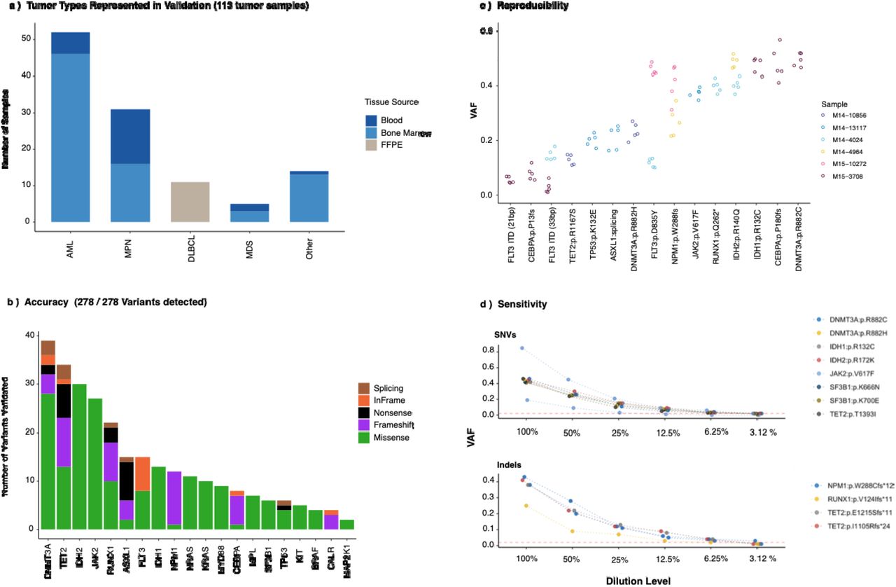
We developed MSK-IMPACT Heme to target 400 genes which are known to be involved in the pathobiology of hematologic neoplasms, are used for diagnosis and prognostication in hematological cancers, and are targets of experimental or approved therapeutic agents (Supplementary Table 1). We have previously described the application of paired tumor normal sequencing for patients with solid tumor malignancies to identify definitive somatic mutations of tumor origin. 6,9–11. To confidently identify somatic mutations in hematologic tumor cells, we used either saliva or nail clippings as a source of germline DNA since genomic material from whole blood may contain high levels of contaminating tumor cells and would not be suitable as a source of both tumor and germline mutations (Figure 1A, see Methods for details). During the analytical validation, mutation detection demonstrated 100% sensitivity and 100% specificity for 278 known mutations in 113 samples across a range of allele frequencies (range: 0.02-0.97) (eFigure 1). Following approval from New York State Department of Health (NYS-DOH), between December 2016 and August 2019, we sequenced 2,383 tumor samples, from 1,937 patients, representing 85 different hematological malignancies (Figure 1B). Of the 2,383 sequenced tumor samples, 1,603 (68%) were sequenced with matched nail DNA, 664 (28%) with matched saliva, and 27 (1%) with both. For the 67 (3%) samples, from 48 patients, that were sequenced following allogeneic stem cell transplantation, both host and donor DNA derived from non-neoplastic tissues were available at the time of sequencing (Figure 1D).

a)Tumor and tissue types sequenced for MSK-IMPACT Heme assay validation. b) accuracy, c) reproducibility, and d) sensitivity results.

a) Overview of the MSK-IMPACT Heme clinical workflow. Distribution of b) tumor types profiled by MSK-IMPACT Heme including c) patient sex by tumor type, and d) patient matched normal sample type used for analysis. e) Variant allele frequency (VAF) of somatic mutations in tumor and matched normal tissues. f) Heatmap showing the percentage of patients with tumor somatic mutations observed in matched nail or saliva tissues. The first number in each cell indicates the number of patients where a tumor mutation is observed in the normal comparator, and the second number indicates the total number of patients profiled with the corresponding. g) Heatmap showing the distribution of genes, where a somatic mutation is found in the tumor and the rate of observing the same variant in the matched normal, indicated with the color-scale. Gray cells indicate that for a given tumor type, either a somatic alteration was not detected in the tumor in that gene or the matched normal sample type (nail or saliva, shown above the heatmap) was not sequenced.
We observed somatic tumor mutations in both saliva and nail at different levels based on disease modality (Figure 1E). While nail DNA was most often purely germline, contaminating tumor DNA was observed with a VAF > 2% in 117 of 1,295 (9%) patients for which nail DNA was sequenced and enriched in chronic myeloid neoplasms, such as a myeloproliferative neoplasms (MPN, PMF, ET, and PV, 43 out of 170 patients, 25.3%), MDS (25 out of 132 patients, 19%), CMML (n=5 of 21 patients, 24%), and AML (n = 16 out of 170 patients, 9%). (Figure 1F). Of the 16 AML cases, the majority arose from an antecedent chronic condition or exposure including prior myeloid neoplasm (n=5), prior chemotherapy/radiation exposure (n=3), or prior cytopenia suggestive of a myeloid neoplasm (n=2). Despite the presence of contamination, the variants detected in nail samples were found with high tumor:nail VAF ratios in virtually all cases (median 8; range 1.5 - 38), supporting the utility of nail control samples towards deciphering the germline versus somatic nature of variants detected in neoplastic patient samples. Somatic variants were rarely identified with VAFs >=10% in nail samples and were primarily confined to disease-defining alterations associated with loss of heterozygosity (LOH) in the tumor sample, such as JAK2 and TET2 in myeloproliferative neoplasms. These alterations were still easily identified as somatic variants owing to the retention of high tumor:nail VAF ratios (Figure 1E). We detected 59 variants with a VAF > 2% in saliva controls from 31 patients, with the vast majority diagnosed with lymphoid neoplasms (90%). The most frequently identified mutations were in DNMT3A, TET2, and TP53, which are commonly associated with clonal hematopoiesis and suggest the presence of a concurrent clonal myeloid process. While only a negligible number of patients with myeloid malignancies (n= 6/1,026) were sequenced with a saliva normal comparator, these saliva controls contained high levels of contaminating tumor DNA, up to 38% VAF (Figure 1 E-G). This finding is consistent with other studies 12, which suggest a limited role for saliva as a germline control in myeloid neoplasms. (Figure 1 E,G).
To highlight the importance of sequencing a matched germline comparator, we analyzed variant calls made in all targeted exonic regions of the MSK-IMPACT Heme panel resulting from ‘unmatched’ variant calling of these tumor samples against a pooled control sample composed of ten diploid blood samples. This analysis resulted in 48,248 variants that were properly filtered by the matched tumor-normal analysis pipeline, but otherwise passed criteria for clinical reporting, namely minimum VAF (0.05), variant sequence reads (10) and their absence from a panel of 25 curated normal samples, known to be lacking any hematologic malignancy. Of these, 27,611 (57%) were present in the gnomAD database with any population frequency >0.01, the primary recommendation for population database filtering from the joint consensus of AMP, ASCO, and CAP pathologists 13, and therefore annotated as putative germline variants that could be dropped in a tumor only analysis. Of the remaining 20,637 putative germline variants, only 928 were within 45% - 55% VAF range and 9,157 (44%) were present in COSMIC v94 database, present in 2,271 tumor samples, or 95% of our sequenced cohort. These represent germline variants that would have been incorrectly reported as somatic in an unmatched analysis, with potential adverse clinical implications. For instance, while specific mutations are not required in the FDA approval for hypomethylating agents (HMAs) in myeloid neoplasms, their presence has been associated with response to HMA treatment, and inaccurate reporting could alter choice of therapy.14 In this analysis we identified a total of 83 germline variants from myeloid neoplasms in genes associated with response to HMA treatment that could have been misattributed to being of somatic origin without a matched normal sample: TET2 (n=47), IDH2 (n=19), IDH1(n=10), DNMT3A (n=4), and TP53 (n=3) (Supplemental Table 2). In addition, the persistence of somatic alterations is often used to monitor response to therapy, therefore misattribution of these alterations as somatic could lead to inaccurate monitoring results in followup samples.
Further, to identify prognostically and diagnostically important small and large scale somatic copy number alterations (SCNAs), we developed a novel algorithm (FACETS2n), which leverages coverage data from patient unmatched normal samples and combines with patient matched allele frequencies to estimate integer level copy number values as well as allelic imbalances such as copy-neutral loss of heterozygosity (CN-LOH) (see Methods). A comparison of results from high-density single nucleotide polymorphism (SNP) array and MSK-IMPACT Heme FACETS2n analysis from 64 clinical samples showed highly concordant results with 92.9% sensitivity and 100% specificity. Discordant calls between SNP array and FACETS2n were attributed to sub-clonal calls made by SNP array and/or tumor fractions below the sensitivity of FACETS2n (less than 20%) as determined by serial dilution of a well characterized tumor sample. (Supplemental Table 3-5).
Having established saliva and nail tissues as suitable controls for identifying true somatic SNVs and indels, we sought to further leverage paired tumor-normal matched sequencing data to assess the allele specific copy number alterations of this cohort. The identification of somatic copy number alterations (SCNAs), including gains and losses in chromosomal arms, has both diagnostic and prognostic implications for hematologic malignancies. 15–17 Historically, karyotype, fluorescent in-situ hybridization (FISH), and single nucleotide polymorphism (SNP) arrays have been used to detect clinically relevant SCNAs. The application of the FACETS2n algorithm to these sequencing data allowed the identification of focal amplifications and deletions as well as broad chromosomal arm level gains and losses. We detected focal copy number alterations in 854 patients (44.1%) whereas 1,146 patients (59.2%) had a chromosome arm level copy number alteration detected. The identification of SNVs, indels and SCNAs in a single assay afforded efficiencies in tissue management and the ability to provide clinically actionable results from a single assay in a clinical setting.
Use of host and donor normal controls to identify somatic alterations in the transplant setting
Confident identification of somatic variants in samples from relapsed patients in the post-transplant setting is a distinct challenge. By sequencing donor derived DNA, we were able to confidently identify and remove donor germline polymorphisms in 47 out of 48 patients profiled following transplant. For one patient, a TP53 variant identified post transplant was also detected in the donor blood sample, but it was not possible to distinguish the germline vs somatic nature based on VAF alone in the tumor sample. In a second patient we identified a putative donor derived somatic variant, DNMT3A p.R882C, likely of clonal hematopoiesis origin. To further demonstrate the utility of a unified analysis using both host and donor normal tissues, we present the case of a 37 year old female who underwent allogeneic stem cell transplantation from an HLA matched unrelated donor for treatment of acute myeloid leukemia. A bone marrow biopsy was performed on day 98 post-transplant for assessment of suspected relapse, which was confirmed with 56% myeloblasts. Engraftment assessment by STR showed a chimeric status with 56% host component (eFigure 2A). We performed MSK-IMPACT Heme on this relapse bone marrow using a pooled control sample as a comparator and called variants. To distinguish somatic mutations from germline polymorphisms, all variant calls were genotyped in the host nail and donor blood samples. Somatic mutations were defined as those with a variant allele fraction (VAF) of at least 0.02 in the bone marrow and not detected in host and donor samples. This approach allowed us to accurately distinguish all host and donor polymorphisms from somatic mutations in this chimeric patient where the range in VAF of host and donor derived polymorphisms overlapped that of the true somatic mutations (range = 0.13 - 0.22) (Figure 2A).
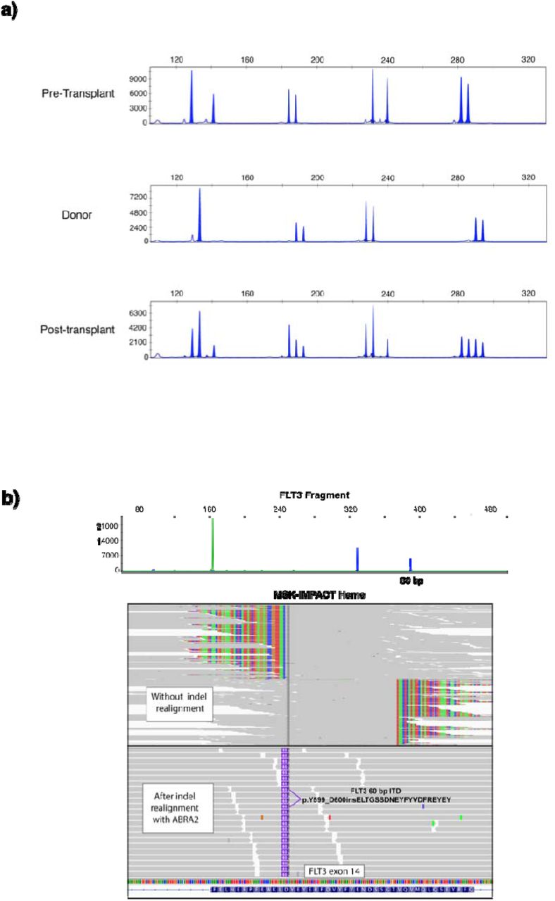
(a) STR analysis of pre and post transplant patient bone marrow and donor blood samples (b) FLT3 fragment analysis (top) and indel realignment with MSK-IMPACT Heme are concordant for identification of a 60bp ITD.
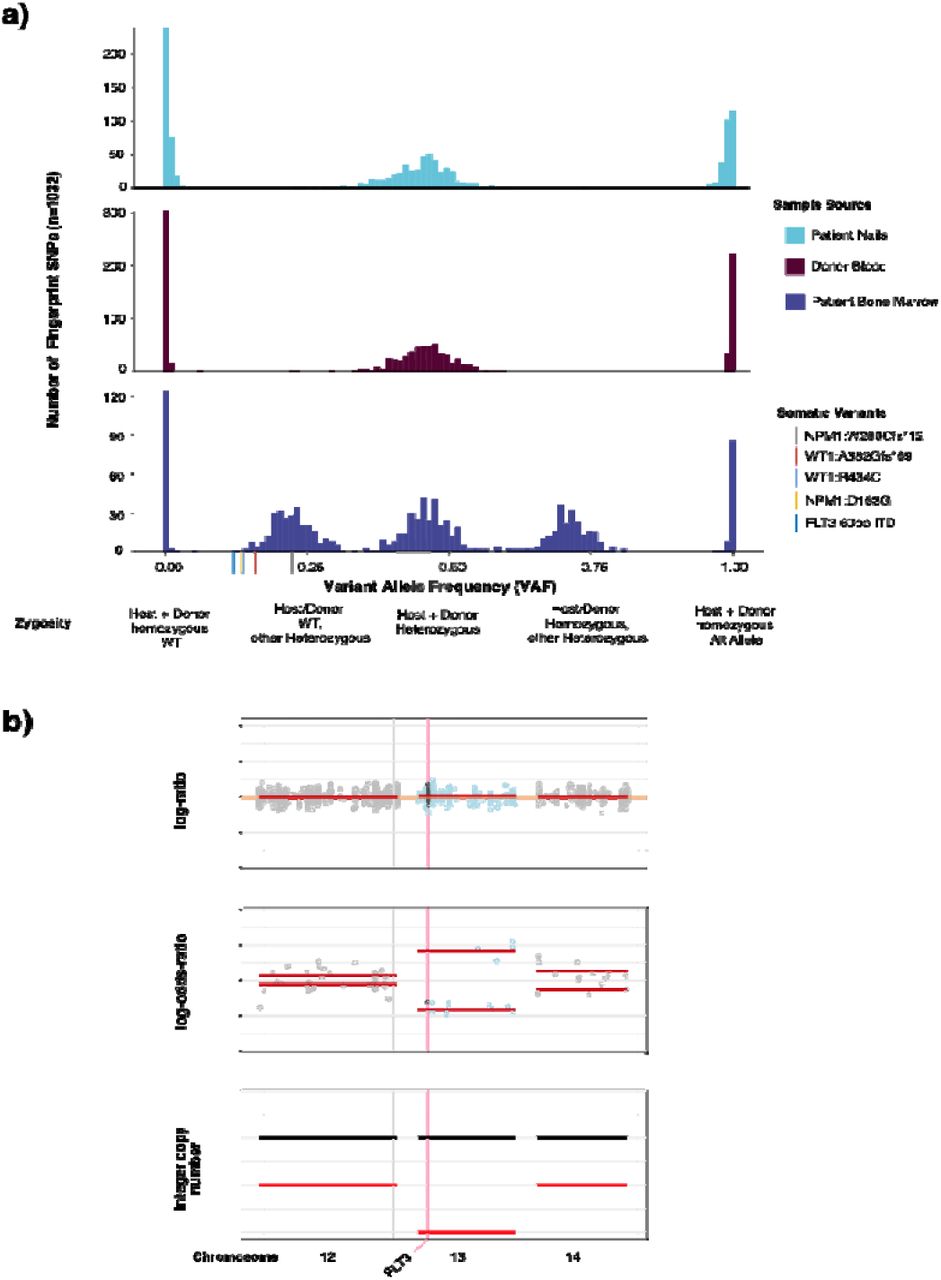
Example patient case highlighting ability of IMPACT-HEME and donor/host matching to detect complex mutations and allele-specific copy number in a post-transplant chimeric patient. (a) The distribution of VAF of somatic mutations, host SNPs, and donor SNPs. (d) Detection of CN-LOH of chromosome 13q, including FLT3.
In addition to removing background polymorphisms, the use of FACETS2n enables more sophisticated local copy number analysis in the post-transplant setting. While computational methods have been developed to infer CN-LOH from SNP array data, both with and without an appropriate matched normal, these methods are impeded by false positives when using unmatched normals 18,19 and have not been optimized to analyze not applicable to samples from patients following allogeneic stem cell transplant chimeric patients due to the potential presence of heterozygous SNPs from more than one individual and unchanged integer copy number. To deal with these challenges, we adapted the FACETS 20 algorithm to use the intersection of heterozygous SNPs between baseline host and donor(s) samples to calculate variant allele log odds ratios with the post-transplant sample and determine regions of allelic imbalance genome wide. To illustrate the power of this approach, we present the case of a patient with a history of AML with a FLT3 internal tandem duplication (ITD) mutation who underwent allogeneic stem cell transplant. FLT3 ITD mutations, such as the 60bp FLT3-ITD detected in this bone marrow (See Methods), are recurrent somatic alterations in AML and typically detected using PCR and capillary electrophoresis assays. (eFigure 2B). Using DNA derived from patient nails and donor blood as baseline sample comparators to the post-transplant bone marrow biopsy, we were able to detect CN-LOH of chromosome 13q (Figure 2B), indicating loss of the wildtype (WT) FLT3 allele. This case illustrates the power of the joint utilization of matched patient and donor normal tissues to differentiate between somatic alterations and both host and donor derived common polymorphisms, as well as to identify allele specific copy number changes in patients after transplant.
Profiling of sorted aberrant cell populations to increase diagnostic accuracy
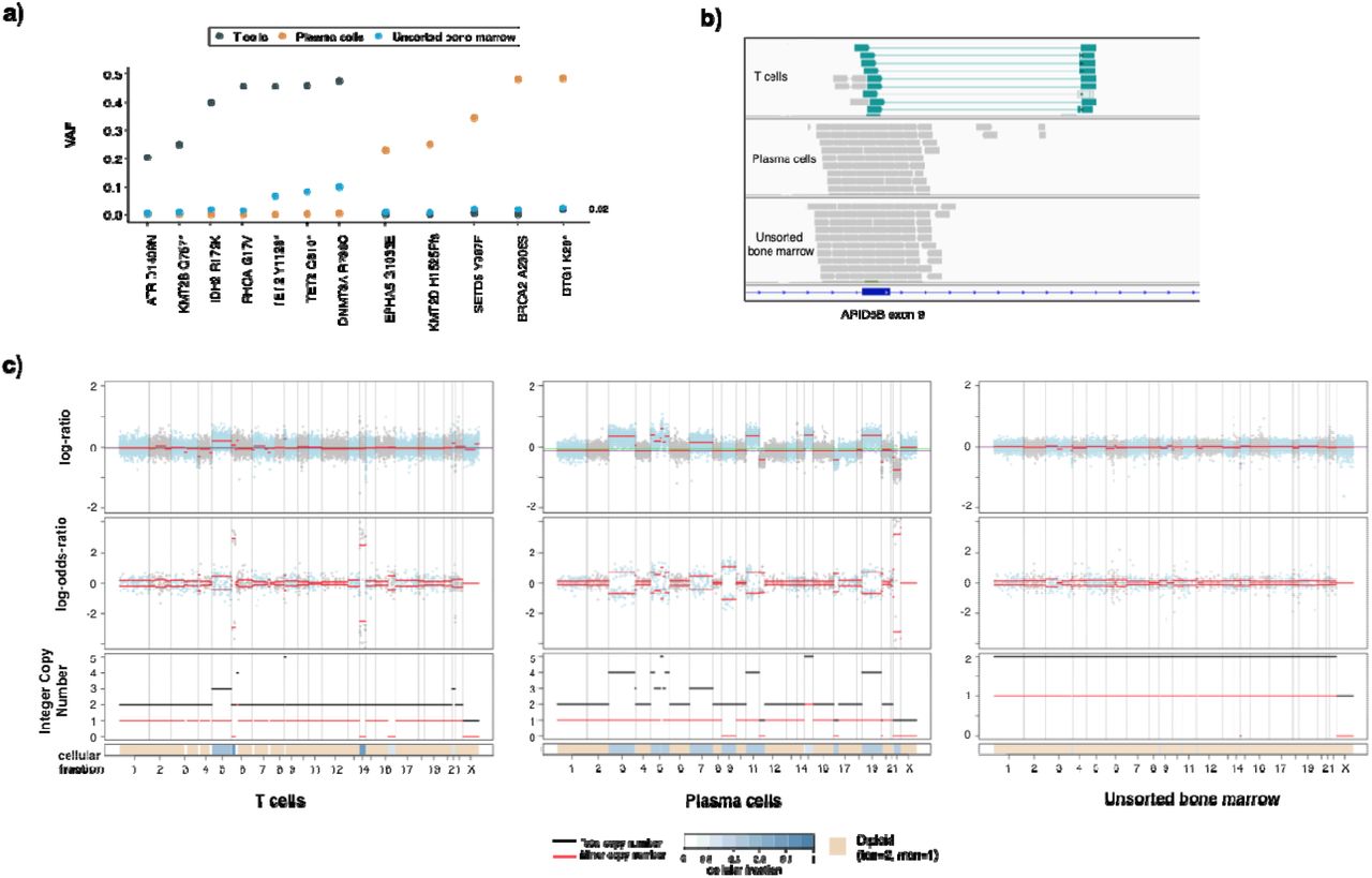
The presence of multiple atypical or neoplastic populations in a sample is not uncommon in patients with hematologic malignancies. These may form part of a clonally heterogenous, single neoplastic process or may represent multiple synchronous neoplastic clones. Clinically, this difference is often difficult to tease out and patients may remain under- or mis-diagnosed and mismanaged. The use of flow sorting or other enrichment practices is a highly valuable approach, and may be successfully performed to enrich very small populations for downstream analysis with our hybridization capture assay. To demonstrate the utility of analyzing flow sorted samples with MSK-IMPACT Heme, we highlight the case of a 72 year old male undergoing diagnostic workup for angioimmunoblastic T cell lymphoma (AITL). Morphologic and immunophenotypic assessment of a bone marrow sample demonstrated low-level involvement by AITL (<5% by CD3/PD-1 immunohistochemistry) and the concurrent presence of a clonal plasma cell population, which accounted for 15% of cells on the aspirate smear and 1.9% of WBC by flow cytometry. Although clonal plasmacytosis has been reported in AITL, 21,22 it remained unclear whether this represented a secondary neoplasm or a reactive expansion. Abnormal T cells and plasma cells were therefore sorted by flow cytometry and submitted for mutational analysis. Independent molecular profiling confirmed the two populations had distinct mutational profiles with the T cell population harboring IDH2, RHOA, DNMT3A, and TET2 mutations 23,24, typical of AITL, while the plasma cells harbored BRCA2, BTG, EPHA5, KMT2D, and SETD5 mutations (Figure 3A). In addition, the two samples harbored unique copy number alteration profiles supporting the diagnosis of 2 separate neoplasms (Figure 3B,C). Of note, only the DNMT3A and TET2 mutations were identified in the unsorted marrow, suggesting that other mutations in the subpopulations were masked as an overall dilution effect in the bulk sample. While DNMT3A and TET2 mutations have been reported to reside in both AITL and clonally related CH25, which may account for the detection of these alterations in both the enriched T-cell and unsorted samples, 26,27 the ability to sort and enrich samples is a powerful tool to interrogate mixed hematopoietic samples to assess clonal relatedness and understand the underlying biology of each population.

Example case of utility for flow-sorted genomic analysis in a patient with AITL and an atypical plasma cell population. a) Somatic mutational profiles of T cells, plasma cells and unsorted bone marrow highlighting distinct mutation patterns in different populations. b) ARID5B structural rearrangement (inversion) detected only in the T cell population. c) The unique somatic copy number alteration profiles of sorted T cells and plasma cells compared to unsorted bone marrow further support that this populations are clonally distinct.
Somatic Genomic Landscape
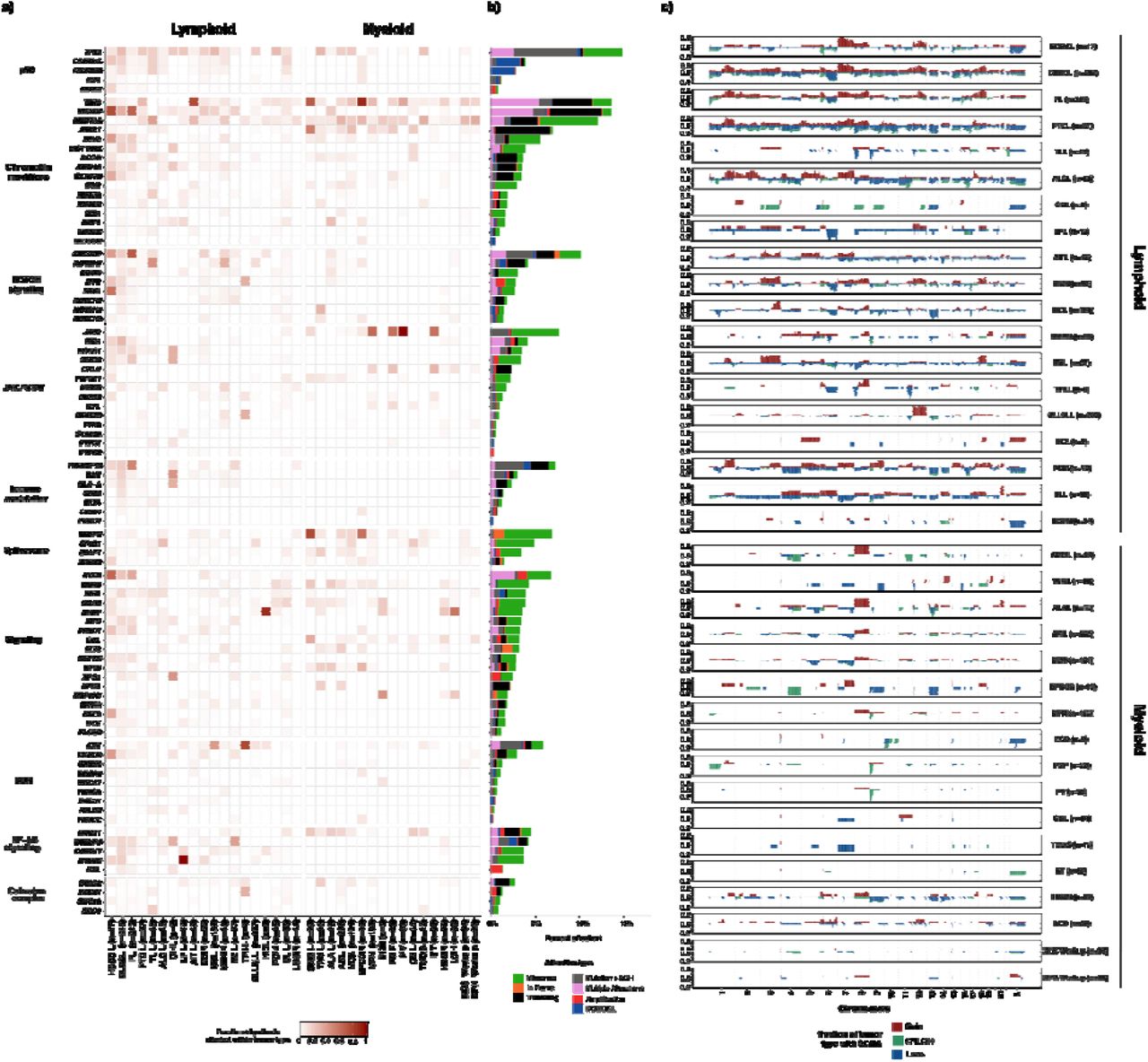
We identified 12,893 somatic mutations, 4,231 gene level and 7,566 broad chromosome arm level somatic copy number alterations from 2,290 samples. Implementation of the MSK-IMPACT Heme workflow enabled the characterization of complex tumor specimens, including flow sorted samples and tumor samples from chimeric post-transplant patients. Somatic genomic alterations including nonsynonymous SNVs, indels, focal and chromosome arm level copy number alterations were identified in 1,885 of 1,937 patients (97.3%). A total of 1804 patients (93.1%) had at least one SNV or Indel identified (median=4, range 0-191). The most commonly identified SNVs were in KMT2D (n= 291, 15%), TP53 (n=288, 15%), TET2 (n=254, 13%) and CREBBP (n=216, 11%). (Figure 4A) We observed broad, tumor purity corrected chromosome level alterations more commonly in lymphoid malignancies (69%, n = 932 / 1357) compared to myeloid neoplasms (37%, n = 377 / 1026; p < 0.001, Fisher’s exact test). The most prevalent arm level SCNAs in lymphoid neoplasms were +7p (n=157, 12%), +18q (n=153, 11%), +12q (n=148, 11%), del 17p (n=215, 16%), del 6q (n=196, 14%), and del 13q (n=169, 13%). For myeloid neoplasms, trisomy 8 (n=63, 6%), +21q (n=27, 3%) and +1q (n=18, 2%) were the most prevalent broad gains, while del 7q (n=59, 6%), del 17p (n=42, 4%), del 5q (n= 40, 4%), and del9p (n=40, 4%) were the most common broad chromosomal losses (Figure 4C). These findings have been well described in myeloid neoplasia and in particular del5q and del7q are considered sufficient to render a diagnosis of MDS, even in the absence of morphologic dysplasia.16 We further compared biological pathways based on the genes in these deleted regions. Lymphoid neoplasms were significantly enriched for deletions in genes of following pathways: p53 (18% vs 5%, q=1.15×10⁻¹⁵), immune modulation (11% vs 0%, q=9.25×10⁻¹⁸), NOTCH signaling (10% vs 2%, q=1.47×10⁻¹⁰), chromatin modifiers (9% vs 5%, q=4.18×10⁻²), DNA damage response (7% vs 0%, q=9.51×10⁻¹²), and NF-kB signaling (6% vs 1%, q=2.26×10⁻⁷). The most prevalent focal copy number alterations in myeloid neoplasms were deletions of TP53 (n=42, 5%), JAK2 (n=44, 5%), FLT3 (n=17, 2%), TET2 (n=16, 2%), and EZH2 (n=16, 2%).

Somatic genomic landscape of hematologic malignancies profiled by MSK-IMPACT HEME a) Recurrent somatic alterations across common tumor types and pathways in lymphoid and myeloid neoplasms b) Bars indicate the percentage of cases harboring different classes of genomic alterations, with integration of mutation and allele specific copy number status. c) Genome wide somatic copy number (SCNA) profiles in main tumor types.
In addition to gene level copy number alterations, FACETS2n enables accurate assessment of allele-specific copy number state, including copy neutral loss of heterozygosity (CN-LOH). CN-LOH was identified in 433 samples (19%) and, similar to global copy number changes, was more frequently noted in lymphoid neoplasms (n= 294/1357, 22%) compared to myeloid (n=139/1026, 14%, p<0.001, Fisher’s exact test) including FL (n=103, 48%), DLBCL (n= 92, 43%), and HGBCL (n=6, 35%). In myeloid malignancies CN-LOH was observed in acute leukemias including AML (n=36) and BLL (n=6), or chronic myeloid neoplasms including PMF (n=14, 50%), CMML (n=9, 35%), and PV (n=13, 34%). The most frequent chromosome arm level CN-LOH events were identified in 6p (n=81), 9p (n=65), 16p (n=50), 9q (n=48), 16q (n=46), 17q (n=46), 15q (n=44), 19p (n=44), 13q (n=43), and 17p (n=41). Interestingly, CN-LOH has been shown to be a mechanism of HLA Class 1 loss in cancer and may underlie the 6p aberrations noted here. 28
Through the integration of SNV/Indel variants and SCNAs, several genes were identified to harbor one mutated allele in conjunction with LOH of the wild type allele. This phenomenon has been well-documented to occur with TP53 across tumor types, ATM in lymphoid neoplasms, JAK2 in myeloproliferative neoplasms, and TET2 in myeloid neoplasms. 29–36. We found similar results with these genes (TP53 n=153, ATM n=53, JAK2 n=33, and TET2 n=30) as well as several other genes. In particular, in FL and DLBCL, the following genes frequently had compound heterozygosity with a mutation and LOH: TNFRSF14 (n=76), CREBBP (n=71), TNFAIP3 (n=30), and B2M (n=29). We also identified genes which harbored multiple somatic variants in a single neoplastic sample, which may reflect bi-allelic inactivation, multiple subclones, or aberrant somatic hypermutation. In myeloid malignancies, multiple alterations were noted in TET2 (n=142, including 24/43 or 56% of AITL samples) and DNMT3A (n=35), while in mantle cell lymphoma, ATM (n=14, 10%) frequently harbored multiple mutations. FL and DLBCL showed multiple mutations in the same patient of KMTD (n=101), CREBBP (n=39), and HIST1H1E (n=20), in addition to aberrant somatic hypermutation of BCL2 (n=58), PIM1 (n=41), and SOCS1 (n=22). TP53 harbored multiple mutations across lymphoid and myeloid malignancies (n=82) (Figure 4B).
Mutational Signatures
The application of DNA sequencing in conjunction with advances in mathematical models have aided the discovery and understanding of the mutational processes that underlie the acquired somatic variants of cancer genomes. 37–39. In clinical tumor profiling, the deciphering of mutational signatures can aid diagnosis, disease prognosis, and treatment decisions. 40–43 However, identification of mutational signatures have occurred mostly in solid tumor cohorts, mainly due to the lower levels of somatic mutation in blood cancers relative to solid tumors 37,44. In the MSK-IMPACT Heme cohort, we calculated the tumor mutation burden (TMB, see Methods) for all samples (range 0-192.9, median 3.7 mut/Mb) (Figure 5A). Relative to myeloid malignancies, lymphoid tumors were characterized by a higher TMB (mean 8.7 vs 3.0 mut/Mb, p<0.001). For the 261 tumors (11%) with elevated tumor mutation burden (>12.9 Mut/Mb, see Methods), all synonymous and nonsynonymous single nucleotide variants were decomposed into COSMIC v3.1 SBS signatures with the inclusion of recently described MMRd signatures as previously described 39,45 (See Methods).

a) Prevalence of somatic mutations across main tumor types and the mutational spectra b) Mutational signatures, sorted by dominant signature for the 261 tumors with elevated mutation burden (>12.9 Mut/Mb)
We identified tumors with mutational processes attributable to activation-induced cytidine deaminase (AID) activity, DNA polymerase eta, mismatch repair (MMR) deficiency, exposure to ultraviolet light (UV), chemotherapy treatment, apolipoprotein B editing complex (APOBEC), and clock-like mutational processes (Figure 5 B). Mature B-cell neoplasms with elevated tumor mutation burden (n=231) displayed dominant mutational signatures associated with genome instability as mediated by AID and the error-prone DNA polymerase eta in conjunction with clock-like mutational processes.46–48 We observed ultraviolet light exposure as a dominant signature in cutaneous T-cell lymphomas (n=11) in addition to DLBCL tumors from two patients, for which clinical histories indicated that these two tumors likely originated near the skin.
Nine tumors from seven patients exhibited a dominant MMR signature, including all four relapse BLL tumor samples with elevated tumor mutation burden. Of the nine tumors with MMR signatures, only one DLBCL sample did not have a clear alteration in the MMR pathway. This case had a lower TMB (20 Mut/Mb) and estimated tumor purity (28%) relative to other MMR tumors and only 43% of mutations attributed to MMR signature. Other signatures attributed to the mutational profile of the tumor were associated with AID, polymerase eta, and clock-like mutational processes. We also observed somatic MLH1 alterations in the two BLL samples with highest mutation burden concurrent with MSH6 frameshift variants and heterozygous loss of MSH2/6. In the first sample, we detected CN-LOH of MLH1, and in the other, a splice variant (c.790+1G>A) previously reported to result in exon 9-10 skipping and reported as a pathogenic germline variant in many individuals with a family history of Lynch-syndrome associated tumors exhibiting microsatellite instability 49,50. No somatic PMS2 alterations were detected in tumors with dominant MMR signature. Both AML samples with elevated mutation burden had dominant mutational signatures for chemotherapy that corresponded with their treatment histories. The two samples with elevated mutation burden and a dominant APOBEC signature were plasma cell myelomas, which has been previously shown to be a poor prognostic indicator 51. Taken together, we show the ability of mutation signature analysis from targeted sequencing of hematologic cancers with elevated TMB to identify underlying mutational processes, with potential to impact patient management using these data.
Clinical Actionability
We also sought to assess the clinical utility of prospective molecular profiling to guide patient management using OncoKB (http://oncokb.org), an expert curated precision oncology knowledge base. OncoKB annotates the oncogenic effect and clinical implications of somatic molecular alterations and has recently expanded to include alterations in hematologic malignancies 52,53. Key to OncoKB is its level of evidence system that annotates molecular variants based on the level of evidence that the alteration is either a predictive biomarker of drug sensitivity or important in informing diagnosis or prognostication. By classifying patient samples by the highest level of evidence assigned to detected variants in that sample, we found that 10.6% of patients profiled had at least one potential clinically actionable alteration, defined as carrying ≥1 alterations assigned an OncoKB level of evidence 1-3B. 54. (Figure 6A). Moreover, 43% of patients had at least one alteration with a diagnostic (Dx) or prognostic (Px) significance as defined by the OncoKB Dx and Px levels of evidence 53 or SCNAs detected by IMPACT-Heme meeting IPSS-R criteria or prognostic indicators in CLL (del13q, trisomy 12, del11q, and del17p). Of note, this analysis reflects an underestimate of actionability at a disease level in this cohort, as the MSK-IMPACT Heme assay does not include targets for the detection of actionable gene fusions and may be addressed in future panel design iterations. Instead, transcript fusion detection is accomplished by a companion RNA based NGS assay 55.

(a) Percentage of samples across all tumor types that harbor a mutation considered clinically actionable according to the OncoKB therapeutic levels of evidence. (b) Distribution of IPSS-M algorithm calculated scores and risk categories identified in the 101 cases in the MSK-IMPACT Heme cohort. (c) The number of patients in the MSK-IMPACT Heme MDS cohort with the given genomic alteration and their stratification into IPSS-M risk categories.
In MDS, the International Prognostic Scoring System–Revised (IPSS-R) is the current standard for patient risk stratification, which relies on clinical parameters of cytopenias, bone marrow blast percentage, and cytogenetic features, but does not consider gene mutations 15. The recently described IPSS-Molecular (IPSS-M) model includes these features in combination with genomic profiling to improve risk stratification (https://mds-risk-model.com) 56. Here, we applied the IPSS-M model to the 101 patients with MDS for whom we also had the required clinical and cytogenetic data to stratify each patient into IPSS-M risk categories. After application of this algorithm, 32.3% (n=33) of cases were classified with a risk category of Very High, 15.8% (n=16) as High, 10.9% (n=11) as Low, and 6.9% (n=7) as Very Low. (Figure 6B). Combining somatic copy number alterations detected by MSK-IMPACT Heme with conventional karyotyping (G-banding) and FISH allowed for a more sensitive detection of copy number alterations, mainly due to the detection of CN-LOH via IMPACT-Heme. Amongst those patients categorized as very high risk, we identified 14 patients with multiple hits to TP53, two patients with multiple TP53 variants and 12 patients with a single variant and loss of heterozygosity. For patients with a single TP53 hit, 36% (n=4) were classified as very high risk. We also demonstrate the ability of IMPACT-Heme to distinguish between subgroups of SF3B1 altered patients, with a single patient identified with mutated SF3B1 and isolated del5q versus 14 patients identified with SF3B1 alpha (lacking co-mutations in BCOR, BCORL1, NRAS, RUNX1, SRSF2, or STAG2) and associated with favorable outcomes 56. (Figure 6C)
DISCUSSION
We herein report the experience of a large institution-wide, prospective clinical sequencing effort to guide the diagnosis, prognosis, and therapy selection for patients across the spectrum of hematologic malignancies. As we previously demonstrated in solid tumors, 6 we now show that this type of enterprise-scale sequencing of neoplastic and matched normal samples is feasible in hematologic cancers, including in patients following stem cell transplant, within a clinical actionable turnaround time. Through this effort, we have generated an extensive collection of manually reviewed mutations and SCNAs in 2,384 samples from 1,937 patients in 85 detailed tumor types. This cross-malignancy dataset will support explorations of driver alterations across all blood cancers to support discovery of rare and unanticipated clinically actionable alterations. With continuing growth in the realm of precision therapeutics, this data set will prove a transformative resource for identifying novel biomarkers to inform prognosis and predict response and resistance to therapy.
In contrast to solid tumors where the primary focus of genomic profiling has been the selection of targeted therapy for key single genetic drivers, the aims of genomic profiling in hematologic cancers are heavily invested on refining a diagnosis and providing prognostic information, with therapy selection often supported by the former. Broad genomic profiling provides a more accurate diagnosis and risk stratification of individual patients at the time of diagnosis and may also predict response and/or outcomes after selected treatments. For instance, TP53 mutations are consistently associated with shorter survival after allogeneic stem cell transplantation and somatic mutations in epigenetic pathways (TET2, IDH1/2, WT1, and DNMT3A) may confer increased sensitivity to hypomethylating agents. 14,57,58 Somatic mutations may require reassessment to update individual risk after treatment, at the time of significant clinical changes or before disease-modifying treatments. Our approach to testing, incorporating routine sequencing of appropriate control samples, enables the unequivocal identification of somatic genetic variants in a way that is scalable even in the context of an allogeneic transplant. It also allows the determination of donor-derived variants which may necessitate monitoring in both the recipient and donor for subsequent development of disease. Additionally, a separate analysis of the normal controls would also facilitate the assessment of key germline events that are relevant to hematologic malignancies.
At the same time, as our understanding of the biology of hematologic malignancies has continued to expand, compounds targeting proteins or signaling pathways disrupted by recurrently mutated genes have become available, notably inhibitors to EZH2 in follicular lymphoma and FLT3, IDH1, and IDH2 in AML. 59,60. One emerging area of study in hematologic cancer is the study of mutation signatures. In solid tumors, mutation signatures such as MMR deficiency and TMB correlate with response to immune checkpoint inhibitors. 61,62 In contrast to solid tumors, hematologic malignancies tend to have lower levels of somatic mutation,37,6 which may account for their relatively disappointing response to immune checkpoint inhibition. 63–67. The unambiguous identification of somatic alterations via the use of a matched normal affords a more accurate assessment of TMB in these neoplasms. Our identification of a small subset of patients with high TMB and/or MMR signatures suggests that these patients could be biological outliers and should be considered for trials of checkpoint inhibition based on these signatures.
In addition to the biological insights and potential for therapeutic targeting afforded by our approach, there is also an opportunity for improved patient monitoring. There has been increasing interest in designing assays for monitoring minimal/measurable residual disease (MRD) following treatment across hematologic malignancy. 68,69 In spite of this interest; however, it is unclear if suitable markers are available for all patients and some guidelines only specify molecular targets for select patients. 70 To address this shortcoming, some groups propose approaches which include use of any somatic alteration as a potential target for monitoring 71,72. These approaches highlight the power of a fully matched sample at initial tumor genotyping and the pitfalls of inaccurate somatic/germline assignment. By removing rare germline variants from reporting through genotyping of candidate variants in matched normal tissue(s), we are able to better identify appropriate markers for MRD assessment and prevent false positive calls.
While this study represents an initial foray into the power of broad scale genomic analysis in hematologic malignancy, additional work remains for the field for clinical genomic analysis to reach its full potential to improve patient care. The best approach to rapidly achieve these goals is through sharing of these datasets across institutions and establishing broad collaborations. To this end, we have deposited our full data set into the cBioPortal for Cancer Genomics (https://www.cbioportal.org/study?id=heme_msk_impact_2022). With continued testing and data sharing, it is our belief that broad genomic assessment will support understanding the pathobiology of, identifying novel drug targets for, and improving noninvasive monitoring for response in all patients with blood cancer.
METHODS
Assay design and validation
We designed custom DNA probes corresponding to all exons of 400 key oncogenes and tumor suppressor genes implicated in hematologic malignancies, including all genes that are targetable by approved and experimental therapies being investigated in clinical trials at our institution.
To determine the accuracy, precision, and sensitivity of the assay, we analyzed DNA from 113 unique tumor DNA samples with known SNVs and Indels in 50 exons of 20 cancer genes previously confirmed by orthogonal methods, as previously described 11. These samples comprised 11 tumor types from blood, bone marrow, and FFPE tissues (eFigure 1A) and had been previously genotyped or sequenced in our clinical laboratory and were confirmed to be positive for mutations by multiple methods. The objective of the accuracy study was to assess the ability of the assay to detect mutations previously confirmed by the reference method in the tested sample. All 278 variants, from 52 exons of 20 genes, were successfully detected with the IMPACT-Heme assay (eFigure 2B). In addition, there was high reproducibility amongst replicates from both inter- and intra-assay experiments. Samples positive for SNVs and indels were tested in triplicate in the same sequencing run and on different days in two additional sequencing pools (eFigure 2C). To determine the analytical sensitivity of the assay, we performed serial dilutions of tumor samples with known variants and determined the VAF at each dilution as output from the variant calling pipeline. The detection limit for low-frequency variants was approximately 2% (eFigure 2D). The ability to detect somatic copy number alterations was demonstrated with samples previously characterized by clinically validated SNP array platforms. MSK-IMPACT Heme was validated and approved for clinical use by the New York State Department of Health Clinical Laboratory Evaluation Program. Following approval, MSK-IMPACT Heme testing was implemented in the clinic to identify genomic alterations that could potentially inform diagnosis and treatment decisions.
Patient consent and accrual
Informed consent for the molecular profiling of patient tumors was obtained under protocol NCT01775072 “Tumor Genomic Profiling in Patients Evaluated for Targeted Cancer Therapy.” The protocol was approved by the Institutional Review Board at Memorial Sloan Kettering Cancer Center and written consent was obtained from all patients. Following consent, either archival or new tumor samples were obtained. The selection of appropriate matched normal was determined after review of patient clinical history and tumor diagnosis. OncoTree (http://www.cbioportal.org/oncotree/), an institutional tumor classification system was used to ensure consistent specimen annotation. Matched saliva was prioritized for lymphoid neoplasms owing to ease of specimen collection and processing and the known paucity of lymphoid components in the samples. Patient matched nail tissue is requested for all myeloid neoplasms due to the high level of neoplastic myeloid cells in patient whole blood and saliva. Patients that had previously undergone hematopoietic stem cell transplantation were sequenced with pre-transplant host and/or donor normal specimens, dependent on engraftment status and tissue availability.
MSK-IMPACT Heme sequencing and analysis workflow
MSK-IMPACT Heme is a custom hybridization capture–based assay for the detection of single nucleotide variants (SNVs), small insertions and deletions (Indels), and somatic copy number alterations. Genomic DNA extraction, library preparation, sequencing, variant calling, and annotation were performed as previously described 6,11. We implemented a custom analysis pipeline (see below) to integrate the analysis of any number of normal samples with a given tumor and provide a reliable assessment of somatic alterations, even in post-transplant chimeric patients. Copy number alterations were assessed using FACETS2n, an allele-specific copy number analysis pipeline for next-generation sequencing data, adapted from the FACETS algorithm 20 to allow the incorporation of multiple normal samples for normalization and determination of allelic imbalance in tumor samples, even those from chimeric patients. All genomic variants called by the analysis pipeline were loaded into MPath, an in-house genomic variant database and user interface that facilitates the manual review of variants and their assessment for therapeutic, diagnostic, and prognostic implications with OncoKB 52. Through the incorporation of variant allele fraction (VAF) in tumor and normal tissues, patient clinical history, and annotated population frequencies, 9 we were able to eliminate variants with low sequencing quality and those of patient and/or donor germline origin.
SNV/Indel calling
Variant calling was performed in paired sample mode using BAM files generated for the tumor sample and the pooled normal control sample processed with each sequencing run. Indel realignment of sequencing reads was performed with ABRA2 73 prior to variant calling to resolve soft-flipped bases to insertions and deletions commonly missed by standard analysis workflows, such as FLT3-ITDs. To the union of calls made by MuTect 74, VarDict 75, and Somatic Indel Detector 76, the genotypes from the patient matched normal sample(s) were incorporated and subjected to automated filtering to generate a complete list of somatic mutation calls, including SNVs and short and long indels. By incorporating the genotype information for patient and donor DNA of non-neoplastic origin, we were able to eliminate variant calls attributed to the germline present in tumor specimens. In detail, all variant calls require a VAF in the tumor ≥ 5 times that of an unmatched normal, a minimum of 20 total reads, 5 alt reads, at least 1% VAF, and presence in less than 20% of our standard normal samples. Furthermore, hotspot sites required an alt allele depth of 8 reads and a VAF ≥ 2%. Non-hotspot sites require a more stringent secondary filtering of at least 10 alt reads, VAF ≥ 5%, and VAF in the matched normal and tumor ≤ 35%. Each alteration identified by the pipeline was annotated with The Ensembl Variant Effect Predictor (VEP) 77 to be compliant with Human Genome Variation Society (HGVS, http://varnomen.hgvs.org) standards and then manually reviewed to ensure that no false positives were reported.
Copy number analysis
Genome-wide total and allele-specific copy number states were calculated for all tumor samples using the open source R package □FACETS2n (v0.3.0). □□ Library specific coverage biases that stem from differences between tumor (FFPE, blood, and bone marrow) and normal (Nails, Saliva, Blood) tissues may result in log ratios with high levels of noise when calculated with matched normal samples. With□FACETS2n, a single unmatched normal is selected from a pool of high-quality normal samples previously processed and sequenced with the MSK IMPACT-Heme assay. These normal samples were selected to have representation of males and females from a variety of tissue types and with different insert size distributions. Selection of a normal sample to be used as a reference diploid genome comparator is performed as previously described 11. The□logOR□of the variant-allele count in tumor versus patient matched normal, an unbiased estimate of allelic copy ratio, was calculated for all heterozygous SNPs (alt allele□freq□between 0.25 and 0.75) in the patient matched normal.□ For patients sequenced following allogeneic stem cell transplantation, logOR was limited to the subset of heterozygous SNPs common to the patient baseline normal sample and all donor samples. For the calculation of integer copy numbers, we utilized a two-pass implementation whereby a low-sensitivity run (cval□= 150) first determines copy number log-ratio corresponding to diploidy. The copy number state of individual genes was determined by a run with higher sensitivity for focal events (cval□= 75).□ The following gene level SCNAs were retained for analysis: amplifications (integer copy number ≥ 5 without whole genome doubling or ≥ 6 with whole genome doubling), homozygous deletions, and heterozygous losses that co-occurred with an SNV or Indel. Broad chromosome arm level copy number gains (integer copy number ≥ 3 without whole genome doubling or ≥ 5 with whole genome doubling) and losses were retained for analysis if they comprised at least 50% of the chromosome arm.
We evaluated the accuracy, sensitivity and reproducibility of MSK-IMPACT Heme in detecting somatic copy number alterations in a validation study of 11 select and clinically relevant regions: chromosomes 3q, 5q, 7q, 8, 11q, 12, 13q, 17p,19, 20q, and the single gene locus TP53. A total of 64 clinical samples were evaluated by both IMPACT-Heme and snp-array, using the results of snp-array as the set of true positive copy number alterations. In these clinical samples, somatic copy number alterations were detected with 92.9% sensitivity and 100% specificity. Three samples with copy number alterations from the validation set were studied over three different sequencing runs for inter-assay reproducibility in addition to being studied in triplicate in the same run for the intra-assay(precision). Concordant results were obtained for all cases in both intra- and inter-assay reproducibility studies. Sensitivity of FACETS2n was evaluated using one FFPE sample from a DLBCL patient with known 12p amplification and 13q loss. Five serial dilutions using DNA from patient matched FFPE normal tissue were prepared (Original, 50%, 25%, 12.5% and 6.25%).
TMB calculation
Tumor mutation burden was calculated as the number of nonsynonymous and synonymous SNVs and Indels per megabase of genome targeted by the MSK-IMPACT Heme panel (1.0837 Mb). We used the distribution of TMB across all tumors to identify highly mutated cases with the formula: median cohort TMB + 2*IQR. Those tumors with a TMB >12.9 Mut/Mb were classified as TMB High and analyzed for mutational signatures.
Mutational signatures
Mutational signatures were assessed for the 261 tumor samples with elevated mutation rates using all synonsymous and nonsynonymous SNVs and SigProfiler software as previously described. 37,39. In order to limit inter signature bleeding that stems from difficult to decipher flat signatures 39,78 and elucidate the mutational processes that contribute to individual cancer genomes, we first performed de novo extraction of single base substitution (SBS) signatures on both individual tumor types and groups of tumors originating from either lymphoid or myeloid lineages. Discovered signatures were decomposed into COSMIC v3.1 SBS signatures with the inclusion of recently described MMRd signatures 45. We then estimated the contribution of each signature to individual cancer genomes using a nonlinear convex optimization programming solver as previously described 39.
IPSS-M
Clinical parameters of percentage bone marrow blasts, hemoglobin levels, and platelet counts were curated for 101 patients with a MDS diagnosis. We then compiled the somatic genomic alterations (SNVs, Indels, SCNAs) and pathologist reviewed assessment of cytogenetic results to derive IPSS-R cytogenetic risk categories, identify complex karyotype, and encode the gene and chromosome level binary variables as input to the IPSS-M algorithm as previously described. 56
Detection of FLT3-ITDs
De novo detection of FLT3 internal tandem duplications (ITDs) using NGS was data was performed by adding an indel realignment step to aligned BAM files using ABRA2 73 which incorporates high quality soft-clipped reads into the generation of contigs that represent variation from the reference genome. ITDs that were resolved via indel realignment were then identified as part of the somatic variant calling pipeline with either the SomaticIndelDetctor and/or VarDict algorithms.
Data availability
The minimal clinical and somatic alteration data necessary to replicate the findings in the article are publicly available on cBioPortal: https://www.cbioportal.org/study?id=heme_msk_impact_2022
Data Availability
All data produced in the present study are available upon reasonable request to the authors
ACKNOWLEDGEMENTS
We gratefully acknowledge Nicole Degroat, Christine England, Sandy Naupari, Jake Bakas, Yingjuan Xu, Hina Patel, Srushti Kakadiya, Jaclyn Rutter, Justina Almodovar, Daviana Martinez-Osorio, Brandon Gray, Hun Jae Jung, Nelio Chaves, Jada Barbee, Kizzia Perez, Aqib Abass, and Shadia Islam for their important contributions. This research was funded in part through the NIH/NCI Cancer Center Support Grant P30 CA008748.
REFERENCES


