Article Text

Statistics from Altmetric.com
The most important diagnostic problem in epileptology is to distinguish epileptic seizures from syncope and from psychogenic attacks. A less common problem is the need to distinguish epilepsy from other paroxysmal disorders with which it may overlap. Improved understanding of ion channel disorders has blurred the definition of epilepsy.1
The diagnosis of episodic altered consciousness rests largely with the clinical history, notwithstanding the remarkable advances in the technology of imaging and neurophysiology. Common reasons for misdiagnosis are:
- ▸
- inadequate or missing history—for example, no witness
- ▸
- clonic movements or incontinence accompanying syncope or psychogenic attacks
- ▸
- undeserved emphasis on a family history of epilepsy or a history of febrile convulsions
- ▸
- overinterpretation of minor electroencephalography (EEG) abnormalities or normal age specific variants.
SYNCOPE
Syncope is the most common “non-epileptic” cause of altered consciousness. The two main types are reflex (vasovagal) and orthostatic syncope. Less common but more serious causes include cardiac and central nervous system syncope.
Reflex (vasovagal) syncope
This is caused by exaggerated but normal cardiovascular reflexes, and so occurs in otherwise healthy individuals, mainly children and young adults.
Clinical features
Table 1 shows characteristics distinguishing vasovagal syncope from epileptic seizures.
Clinical distinction of reflex (vasovagal) syncope from seizures
Triggers include prolonged standing (school assembly), rising from lying (bathroom at night), hot crowded environments (restaurant), emotional trauma, and pain (doctor's surgery). Prodromal symptoms (presyncope) developing over 1–5 minutes include light headedness, nausea, sweating, palpitation, greying or blacking of vision, muffled hearing, and feeling distant.
A witness may describe pallor, sweating, and cold skin. Muscle tone is flaccid sometimes with a few uncoordinated clonic jerks occurring after the fall. A common error is to label syncope as a seizure, given a witness account of shaking (convulsive syncope).2 Incontinence and injury are uncommon and lateral tongue biting rare. Recovery is usually rapid and without confusion.
Mechanism
The mechanism is complex. Venous return falls when blood pools in either the legs (prolonged standing) or the splanchnic vessels (increased vagal tone—for example, response to seeing blood). Cardiac output therefore falls, but the blood pressure is maintained initially by sympathetically induced peripheral vasoconstriction. The Bezold-Jarisch reflex is initiated when vigorous cardiac contractions stimulate ventricular wall mechanoreceptors, exciting 5-hydroxytryptamine (5-HT, serotonin) mediated vagal afferents, falsely “suggesting” an overfilled heart. The resulting surge in vagus activity and reduced sympathetic tone, despite the falling blood pressure, set up a vicious cycle of bradycardia, falling peripheral resistance, and abrupt circulatory collapse. In addition, cerebral vasoconstriction preceding the circulatory collapse explains presyncope preceding major falls in blood pressure.
Investigations
The history usually gives the diagnosis. An electrocardiogram (ECG) is indicated if syncope occurs with exercise, when lying, or with palpitation (see cardiac syncope below). Significant falls in postural blood pressure rarely happen in outpatients; indeed, blood pressure usually shows the normal initial rise on standing. Tilt table testing (60° tilt for 45 minutes) (fig 1A) is therefore important if there is diagnostic doubt.3 It is highly sensitive (< 90%) and specific (70%) for a syncopal tendency. Measures to induce syncope—for example, with isoprenaline—provoke syncope more rapidly but with lowered specificity.

Heart rate and blood pressure changes during tilt table testing in (A) vasovagal and (B) orthostatic syncope.
Management
An explanation and postural advice—head down or lie flat at symptom onset, or rise slowly from lying—is often sufficient. Behavioural psychotherapy and systematic desensitisation may help with specific triggers—for example, seeing blood. Medications include β blockers4 to limit vigorous cardiac contractions, salt loading or fludrocortisone to prevent cardiac underfilling, or selective serotonin reuptake inhibitors to modulate vagal brainstem reflexes. Dual chamber cardiac pacing may help in severe (malignant) vasovagal syncope: maintaining the heart rate despite falling blood pressure may not prevent syncope, but allows presyncopal symptoms (and evasive action) to occur.
Orthostatic syncope (autonomic failure)
People with autonomic dysfunction lose their normal vasoconstriction response to a postural fall in blood pressure. Syncope occurs within seconds or minutes of becoming upright, especially when rising and after meals. Unlike in reflex vasovagal syncope, the skin stays warm, the pulse rate is unchanged despite the fall in blood pressure, and sweating is absent.
Autonomic dysfunction may relate to autonomic neuropathy (for example, old age, diabetes, alcohol, amyloidosis) or complex autonomic failure (multiple system atrophy). Medications such as antihypertensives, phenothiazines, tricyclic antidepressants, anti-parkinsonian treatments, and diuretics worsen the problem.
Lying and standing blood pressure (postural drop without pulse rate change) provides the diagnosis. Other autonomic function tests (blood pressure and pulse rate during hyperventilation or Valsalva manoeuvre) and tilt table testing also help (fig 1B). Exclusion of anaemia or hyponatraemia is important.
Management includes withdrawing provoking medications and avoiding certain situations, such as prolonged standing, large meals, and alcohol. Head-up tilt at night may help. Fludrocortisone acetate (50–200 μg daily) is the usual first line medication.
Cardiac syncope
Cardiac syncope results from either rhythm (electrical) or structural (“plumbing”) disorders, and is potentially life threatening. Some sudden unexplained epilepsy deaths are undoubtedly misdiagnosed cardiac syncope. Syncope occurs from any posture (arrhythmogenic syncope is common in bed), during exertion or emotion. Syncope during exercise requires urgent exclusion of a cardiac cause, though most turn out to be reflex vasovagal syncope.
Tachyarrhythmias
Rapid tachycardia reduces cardiac output through incomplete diastolic ventricular filling. Palpitation occurs during and between attacks. Supraventricular tachycardias are common, often benign, and usually not associated with structural heart disease; ventricular tachycardias are more serious, often occurring with heart disease.
Wolff-Parkinson-White syndrome (short PR interval and delta wave) predisposes to “re-entry tachycardias” through an abnormal short circuit, the bundle of Kent. The related Lown-Ganong-Levine syndrome shows a short PR interval but no delta wave.
Inherited long QT syndromes are potassium or chloride channel disorders giving variable refractoriness between adjacent cardiac myofibrils.5 Resulting “micro re-entry” phenomena provoke “torsades de pointes”, a potentially fatal ventricular tachyarrhythmia where the QRS axis rotates repeatedly through 360°. The main genetic syndromes are Romano-Ward (autosomal dominant) and the Jervell and Lange-Neilsen syndrome (autosomal recessive, more severe, and with deafness). Recurrent syncope develops, particularly on rising. Typically patients fall, lie still, and then convulse. The risk of sudden death lends urgency to family screening.
Acquired QT changes predisposing to arrhythmia are common. Shortening occurs with digoxin, hyperthermia, and hypercalcaemia; lengthening occurs with some antiarrhythmics, antihistamines, and with ischaemic heart disease.
Bradyarrhythmias
Chronic bradycardia—for example, complete heart block—reduces cardiac output, impairing the cerebral circulation; patients develop fatigue as well as syncope.
Carotid sinus hypersensitivity is a common cause of sinus arrest and “drop attacks”.6 This is found in 10–20% of people over 60 years, particularly men, of whom about 20% present with syncope attributable to carotid pressure (for example, tight collar, head turning, etc). Diagnostic carotid sinus massage is performed with the patient supine (repeated upright if suspicion is strong), under ECG and blood pressure monitoring. Each carotid is massaged for < 15 seconds (leaving > 15 seconds between sides). Positive responses are either cardioinhibitory (sinus pause > 3 seconds), vasodepressor (blood pressure fall > 50 mm Hg), or mixed. The risks include prolonged asystole, transient or permanent neurological deficit, and sudden death.
Other causes of reflex bradycardic syncope are the Valsalva manoeuvre (for example, cough or trumpeter's syncope), swallowing (often with glossopharyngeal neuralgia), or micturition.
Structural cardiac causes
Left ventricular outflow tract obstruction (aortic stenosis, hypertrophic obstructive cardiomyopathy) and left ventricular underfilling (mitral stenosis, atrial myxoma) reduce stroke volume. Cardiac pump failure (cardiomyopathy, ischaemic heart disease, arrhythmogenic right ventricular dysplasia (ARVD)) also reduces cardiac output. In each case, however, syncope is principally vasovagal, the vigorous ventricular contractions giving increased vagal tone.
ARVD is autosomal dominant and may present as sudden death. The diagnosis is difficult without a family history since ECG abnormalities (inverted T waves in V2–V4) occur in only 70%, and the patchy right ventricular wall fibrosis may be missed on endocardial biopsy.
Central nervous system (CNS) syncope
There are several patterns of CNS syncope:
- ▸
- Intermittent obstructive hydrocephalus (third ventricular cyst, Chiari malformation) typically presents as “pressure” headache building over several seconds before losing consciousness. The potential result is sudden death.
- ▸
- Concussive (immediate post-traumatic) seizures occur within seconds of an acute head injury. They are non-epileptic, show no typical EEG changes, and do not predict later epilepsy.
- ▸
- Autonomic dysreflexia may follow complete spinal cord lesions. Loss of autonomic blood pressure control allows intermittent massive hypertension, sometimes sufficient to alter consciousness.
- ▸
- Diencephalic attacks following diffuse brain insults (head injury, hypoxic encephalopathy) manifest as intermittent hypertension, sweating, tachycardia, and even loss of consciousness.
Psychogenic attacks
Psychogenic non-epileptic attacks as part of panic disorder and dissociative disorder are commonly mislabelled as epilepsy.7 8
Panic disorder
Anxiety and panic disorder may culminate in syncope through hyperventilation induced hypocapnia and resulting cerebral vasoconstriction. Facial and limb tingling may be lateralised, and easily mistaken for transient ischaemic attacks or seizures. Accompanying symptoms include feelings of panic, light headedness, breathlessness, palpitation, chest and throat tightness, blurred vision, and carpopedal spasm.
Difficulty distinguishing panic disorder from epilepsy arises for three reasons. Firstly, panic may resemble seizures—for example, “frightened” epigastric sensations, palpitation and hyperventilation symptoms (see above). Secondly, panic disorder may result from epilepsy. Patients with epilepsy are understandably anxious, concerned by the risk of seizures, injury, and social embarrassment; resulting panic symptoms initiate a vicious cycle of apparently increasing seizure frequency. Thirdly, panic exacerbates epilepsy through hyperventilation, sleep deprivation, and increased alcohol consumption. Worsening of seizures at times of stress is quite consistent with a diagnosis of epilepsy.
Management
An explanation (supported by written material) of the nature and mechanism of the attacks is helpful. Management aims at avoiding provoking or threatening situations, and reducing both anxiety (for example, benzodiazepines or paroxetine) and physical symptoms (for example, β blockers).
Cognitive behaviour therapy focuses upon the patient's tendency to misinterpret bodily sensations (for example, palpitations or tingling) as indicating impending catastrophe. This may be combined with breathing control and relaxation.
Dissociative non-epileptic attack disorder (NEAD)
Dissociative non-epileptic seizures mimicking tonic-clonic seizures (“pseudoseizures”) cause major diagnostic difficulty and carry significant resource implications and potential morbidity if the diagnosis is missed. Dissociative NEAD may comprise up to 50% of supposed status epilepticus.
Clinical features
Dissociative NEAD is characterised by frequent attacks provoked by stressful events, commonly giving doctors an opportunity to witness attacks. Table 2 shows features distinguishing them from seizures.
Clinical distinction of dissociative non-epileptic seizures (“pseudoseizures”) from epileptic seizures
Patients with dissociative NEAD are usually female (8:1), frequently with previous abnormal illness behaviour and multiple hospital admissions, and often have suffered childhood physical and/or sexual abuse. Their attacks typically begin after the age of 10 years, are resistant to medication, and frequently recur, often with “status epilepticus”. “Pseudostatus” should be suspected when treatment resistant convulsions occur repeatedly, without significant underlying brain dysfunction either clinically, or on EEG or brain scan.
Investigation
Interictal EEG is usually normal; postictal EEG does not show the slowing expected following an epileptic seizure. Video EEG monitoring is especially useful in dissociative NEAD. Not only can the attacks be observed and the EEG seen to be normal, but also the recording can be re-played to the patient, allowing discussion of the experience.
Measuring prolactin concentrations before and 20 minutes after the onset of an attack may help. No rise follows non-epileptic seizures whereas significant elevations follow epileptic convulsions, complex partial seizures or syncope.
Management
Diagnostic certainty is ideal, but complete exclusion of epilepsy is rarely possible; some attacks might still be epileptic. It is helpful to explain that the diagnosis is good news, being potentially curable, and to emphasise the unconscious nature of the attacks occurring as a stress response (comparable to control of breathing and heart rate).
Follow up is important, giving opportunity to discuss problems, fears, and memories. Childhood physical or sexual abuse may be disclosed. Relaxation techniques, breathing control, hypnosis, aromatherapy, or cognitive therapy may help. Antiepileptic medication should be withdrawn when the patient is ready.
Outcome
Significant morbidity can result from NEAD, particularly “pseudostatus”. Medication, cannulation, infection, and assisted ventilation are potentially hazardous, and pregnant women with NEAD risk premature intervention and delivery. Even with sympathetic handling and long term follow up, the prognosis is guarded.
Rages
Rages may resemble ictal or postictal behaviours. Epileptic rages (arising frontally) are typically unprovoked, brief, and followed by sleepiness, whereas episodic dyscontrol shows situational provocation of individual attacks.
Migraine
Migraine equivalents (migraine aura without headache) may pose diagnostic challenges.9 10 Visual migraine auras are zigzag, bright, and monochrome, gradually evolving and traversing the visual field. By contrast, epileptic visual hallucinations are unformed or circular (but not zigzags), multicoloured, and often confined to one hemi-field. Somatosensory migraine typically gives unilateral tingling spreading from one body part to another over 15–30 minutes, resolving from the first (for example, hand) before reaching the next (for example, face). By contrast, epileptic sensory symptoms typically spread quickly to involve the entire ipsilateral side, sometimes with convulsion.
Several migraine variants may be confused with epilepsy. Retinal migraine gives recurrent, often very frequent, unilateral visual aura, without headache or systemic upset. Aspirin is usually effective prophylaxis. Basilar artery migraine, usually in children, overlaps with benign occipital epilepsy (visual aura, headache, vomiting), and manifests as prolonged visual blacking, vertigo, diplopia, loss of consciousness, headache, and vomiting. Migraine syncope (of gradual onset) and even migraine coma may complicate familial hemiplegic migraine. Coma may recur but individual attacks have an excellent prognosis.
Other vascular disorders
Transient ischaemic attacks (TIAs) are broadly distinguished from seizures and migraine by their “negative” symptoms (numbness, weakness or transient blindness) and retained consciousness. Tingling, however, may occur in TIA and focal jerking accompanies the severely compromised cerebral blood flow of bilateral carotid stenosis. Also, bilateral visual loss, sometimes for many minutes, occurs in occipital lobe epilepsy. Altered consciousness occurs in TIA only with ischaemia affecting the brainstem or entire cortex (for example, following cardiac arrest).
Transient global amnesia manifests as a transient (several hours) anterograde memory loss with repeated questioning, and sometimes migraine type headache. Presumably, there is transient ischaemia (vasospasm) of mesial temporal structures. It has an excellent prognosis and rarely recurs. Recurrent or unusually brief episodes raise suspicion of temporal lobe epilepsy, particularly transient epileptic amnesia.
Sleep disorders
Excessive somnolence
Obstructive sleep apnoea is common, treatable, and under recognised. Sleep related upper airway collapse causes frequent apnoeas and sleep fragmentation; the consequent excessive daytime somnolence may suggest epilepsy. Furthermore, the sleep deprivation may exacerbate pre-existing epilepsy. Important predisposing factors are obesity, alcohol, and structural upper airway narrowing (including nasal congestion).
Narcolepsy is well known but overdiagnosed. It presents with daytime somnolence and inappropriate sleep—for example, during conversation or meals. Other symptoms include rapid sleep onset, poor quality night time sleep, hypnagogic (just before sleep) hallucinations, and sleep paralysis.
Cataplexy, effectively REM (rapid eye movement) sleep intruding into wakefulness, is part of the narcolepsy syndrome. Laughter or emotion triggers transient axial and facial atonia with dysarthria or mutism, often with facial jerking, but without altered consciousness. It is treated with clomipramine.
Parasomnia
Parasomnias—including confusional arousals, periodic limb movements, rhythmic movement disorder, night terrors, and somnambulism—are non-epileptic but easily confused with seizures. It is recognised that most familial parasomnias are probably examples of autosomal dominant nocturnal frontal lobe epilepsy (ADNFLE).
REM sleep behaviour disorder results from dissociation between REM sleep and axial atonia; the patient acts out dreams. It is usually of elderly onset, and associated with brainstem disease (multiple sclerosis or multiple system atrophy). Self injury may result and clonazepam is usually effective.
Paroxysmal movement disorders and ataxias
Paroxysmal dystonic disorders overlap with epilepsy and usually respond well to anticonvulsants. Paroxysmal kinesigenic dystonia is a familial condition where sudden movement provokes involuntary unilateral dystonic posturing lasting a minute or so. Tonic spasms of multiple sclerosis are involuntary, often painful, unilateral dystonic movements. Paroxysmal nocturnal dystonia is a form of ADNFLE rather than a primary movement disorder; the term has been largely abandoned.
Periodic ataxias are autosomal dominant ion channel disorders manifesting as childhood or adolescent onset attacks resembling seizures. Episodic ataxia/myokymia (type 1), a potassium channelopathy (chromosome 12p), manifests as ataxia and myokymia lasting seconds to minutes. The EEG often shows sharp and slow waves and true epileptic seizures also occur. Acetazolamide responsive autosomal dominant periodic ataxia (type 2) is a calcium channelopathy (chromosome 19p) related to familial hemiplegic migraine. It presents as vertigo, ataxia, and even blackouts. Acetazolamide is usually effective.
Endocrine, metabolic, and toxic causes
Hypoglycaemia, easily diagnosed and readily reversed, must be considered in anyone with undiagnosed altered consciousness. Insulin treated diabetes mellitus is the obvious cause. Insulinoma, though rare, is frequently missed. It presents as recurrent confusion, early morning seizures, and weight gain (frequent sweet drinks). A 72 hour fast induces attacks with low blood glucose and inappropriately high endogenous insulin concentrations. Alcohol, hepatoma, or undeclared exogenous insulin (for example, Munchausen syndrome) also provoke hypoglycaemia. Hypocalcaemia (from hypoparathyroidism, for example) may present as recurrent episodes of tingling, carpopedal spasm, and syncope. Phaeochromocytoma and carcinoid syndrome may present as confusion episodes.
Porphyria rarely causes unprovoked seizures. However, acute confusion and psychosis may mimic epilepsy and antiepileptic medications (for example, carbamazepine or phenytoin) only exacerbate the porphyria. There is often a family history of psychotic attacks or neuropathy.
Summary
- ▸
- A detailed history and witness account are essential for diagnosing and managing episodic altered consciousness
- ▸
- 20% of patients labelled and treated as epilepsy have other causes for apparent blackouts
- ▸
- Syncope and psychogenic attacks are the most common reasons for misdiagnosis of epilepsy
- ▸
- Clinicians must not rush to a label of epilepsy; trials of antiepilepsy treatment are rarely justified
- ▸
- Features useful in distinguishing syncope from seizure are the triggers (for example, upright, bathroom, blood), the prodrome (especially visual blacking), the absence of lateral tongue biting, and the prompt recovery (wakes on the floor not in the ambulance). Incontinence and injury are less helpful
- ▸
- Vasovagal syncope results from autonomic hyper-reflexia; orthostatic syncope results from autonomic failure
- ▸
- Exercise induced syncope, though often vasovagal, requires urgent cardiac investigation
- ▸
- Features useful in distinguishing psychogenic non-epileptic attacks (“pseudoseizures”) from epileptic convulsions are the triggers (frustration, suggestion, in company), duration (often very prolonged), erratic movements (fighting, pelvic thrusting), remaining pink and breathing, resisting eye opening and eye contact, and often prompt recovery
- ▸
- Panic symptoms frequently accompany epilepsy; they may be caused by, may worsen, and may mimic seizures
- ▸
- Perhaps 50% of adults admitted to intensive care for “status epilepticus” have psychogenic attacks and not epilepsy
- ▸
- Frontal lobe seizures are easily misdiagnosed as psychogenic attacks
Alcohol excess may provoke syncope, seizures or coma through direct toxicity, withdrawal or hypoglycaemia. Figure 2 describes a well known historical example of alcohol related attacks diagnosed as epilepsy. Illicit drugs provoke seizures either by direct toxicity (for example, cocaine) or sleep deprivation (for example, amphetamine, including Ecstasy). Deliberate poisoning in non-drug users may present as undiagnosed altered consciousness. “Date rape drugs” such as flunitrazepam (Rohypnol) dissolve rapidly, are colourless and tasteless, and induce a trance-like state with muscle relaxation. Rape victims have difficulty pressing charges owing to amnesia.
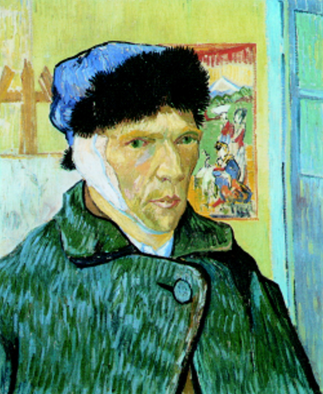
Vincent van Gogh (1853-1890). Self portrait with bandaged ear (1889), courtesy of the Courthauld Institute, London. Van Gogh's “epilepsy” presented as recurrent, prolonged (up to two weeks) attacks of confusion, vertigo, and uncharacteristic violence, followed by remorse and, on one occasion, self mutilation. Suggested explanations have included epileptic psychosis, porphyria, and Ménière's disease. Psychosis induced by absinthe, an alcoholic spirit available in late 19th century France, perhaps exacerbating complex partial seizures, seems the most plausible diagnosis.11
Misdiagnosed epilepsy
Certain epilepsies are commonly initially misdiagnosed as non-epileptic.
Generalised seizures (for example, childhood absences) may be dismissed as poor concentration, with disastrous educational consequences. The diagnostic significance of myoclonic jerks may be overlooked.
Frontal supplementary motor area seizures cause particular diagnostic difficulty. Frequent, brief, and sleep related attacks might suggest parasomnia. Dissociative NEAD may be suspected owing to hyperkinetic behaviour, retained consciousness, and hyperventilation during attacks. Furthermore, seizure activity buried between the cerebral hemispheres may give normal surface EEG (movement artefact only) during seizures.
Medial temporal lobe seizures starting as epigastric “butterflies” can suggest panic disorder; associated palpitation, tachycardia or bradycardia suggest cardiac syncope. Parietal lobe seizures presenting as episodic lateralised sensations or pain without motor components, or occipital seizures manifesting as ictal blindness or visual hallucination, are often misdiagnosed as migraine or psychogenic.
Non-convulsive status epilepticus is easily missed if presenting as acute confusion. Complex partial status epilepticus is the usual cause in adults. Absence status epilepticus is more common in children, though “de novo absence status epilepticus” presents as confusion in adult (usually elderly) patients, sometimes without a history of epilepsy.
Implications for management
A confident diagnosis of epilepsy is essential for its proper management. Where diagnostic doubt remains, clinicians must not rush to a label of epilepsy. An open mind and the passage of time often allow the true nature of the attack to declare itself with fewer problems than accompany a hurried label of epilepsy. Trials of antiepilepsy treatment are best avoided.
References
Key epilepsy references
- 1-1.
- 1-2.
- 1-3.
- 1-4.
- 1-5.
- 1-6.
- 1-7.