Article Text

Abstract
Purpose Simulation techniques in neurosurgical training are becoming more important. The purpose of this study was to determine whether silicone vascular models used in the angiography suite can render improvement in trainee performance and safety in neuroendovascular procedures.
Methods 10 residents from neurosurgery and radiology training programs were asked to perform a diagnostic angiogram on a silicone based vascular model (United Biologics, Tustin, USA). This was done in the angiography suite with the full biplane fluoroscopy machine (Siemens, Munich, Germany). On their first attempt, they were coached by a faculty member trained in endovascular neurosurgery; on their second attempt, they received coaching only if the procedure had stalled. Technique was scored on multiple criteria by the faculty, and total time and fluoroscopy time were recorded on both attempts.
Results In this group of 10 residents, overall procedure time significantly decreased from 51 to 42 min (p=0.01), and total fluoro time significantly decreased from 12 to 9 min (p=0.002) between the first attempt and the second attempt. Technical skill increased significantly in navigation, vessel selection, projection setup, and road map usage.
Conclusions Silicone vascular models used in the angiography suite, with the clinical working tools and biplane fluoroscopy, provide a valuable experience for training residents in diagnostic angiography, and improved performance and safety.
- Technology
- Technique
- Angiography
- Device
Statistics from Altmetric.com
Background
In the course of training neurosurgical residents, residency programs employ many techniques to improve their technical skills and procedural knowledge. In the era of dwindling work hours, many of these techniques must rely on the residents’ own motivations and allow them to train outside of their normal clinical activities which fall under the set time restrictions. Simulators have become one of the many tools utilized to accomplish this goal.
Angiographic simulation allows residents to train and improve procedural skill, either under the direct guidance of a faculty member or on their own time. The distinction between this method of angiographic simulation and other computer simulators previously reported is that this simulator model can be used in the angiography suite in conjunction with the biplane fluoro unit and actual endovascular equipment; it is an accurate physical representation of the vessels of the aortic arch and cerebral circulation. With this model, residents become acclimated both with the catheter and wire techniques and tools, and the manipulation of the angiography machine and framing of the circulation for diagnostic and interventional work.
The use of the physical tools becomes especially valuable for residents on rotation when they are able to use the same moves clinically that they have practiced on the simulator.
Virtual simulators have been shown as feasible methods to improve trainee skills and knowledge when combined with didactic lessons.1 This study was undertaken to determine whether the silicone models in an angiography suite can render improvement in trainee performance and safety. A second objective was to demonstrate the advantages of a silicone model over computer based endovascular simulators which are gaining popularity.
Methods
Ten trainees in neuroradiology and neurosurgery at the University of Wisconsin-Madison were recruited for testing on the simulator. Each trainee received the same baseline teaching from an endovascular neurosurgeon. Trainees were oriented to the silicone model (the Neuro System Trainer; United Biologics, Tustin, USA), the endovascular tools (DAV diagnostic catheter (Cook Medical, Bloomington, USA) and Terumo glidewire (Terumo, Somerset, USA)), and the biplane fluoroscopy unit.
Each trainee wore lead throughout the simulation, and a special low dose fluoro protocol was used. Low dose fluoroscopy and road mapping settings for the Siemens Artis zee biplane angiography system (Siemens, Munich, Germany) were used for training in order to minimize radiation exposure to the operators. Low dose fluoroscopy settings included a reduced frame rate (7.5 fps compared with the standard 10–15 fps) and a lower x-ray dose per frame (29–32 nGy/frame compared with 36–55 nGy/frame for standard and high dose fluoroscopy).
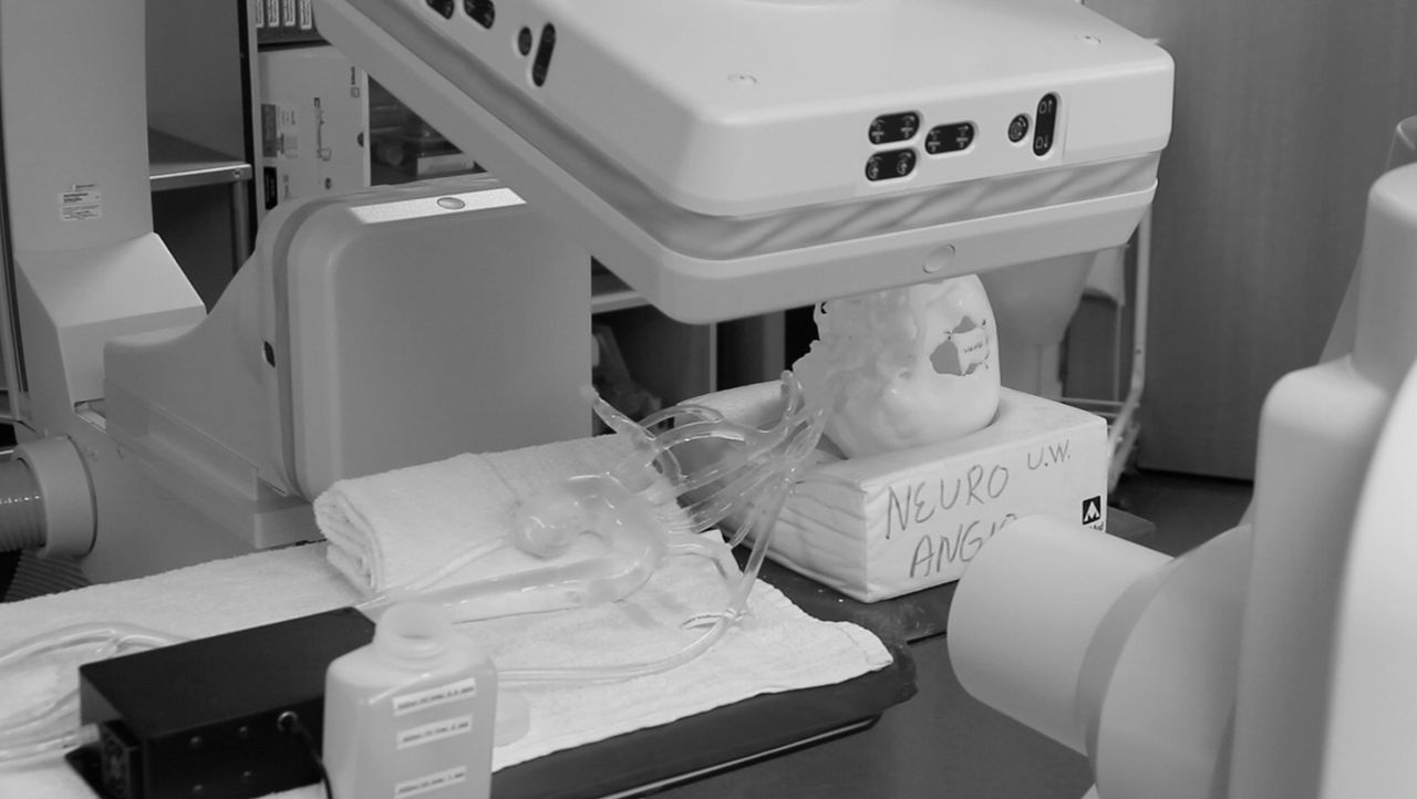
The model is made from silicone and extends from the femoral arteries to the pericallosals and M2 and A2 segments (figure 1). The vessel size is physiologically accurate, and was used embedded in a skull model so that the appropriate angiographic views could be practiced. The vascular model is radiopaque, which allows it to be used either without contrast and visualized on the native, or with contrast and visualized on the subtracted images in the usual way. There is a pump that circulates saline in the model to simulate blood flow. Vessel access was simulated with a rotating hemostatic valve attached to one of the luer lock access points on the model.

The silicone vascular model displayed on the angiography table with the biplane fluoro machine in place. The vascular model is embedded in a skull model to allow the resident to practice acquiring the proper angiographic projections.
Outside of the angiography suite, each trainee received fundamental lessons on how to perform a complete four vessel diagnostic angiogram with the real diagnostic equipment provided. These lessons included instruction in achieving the appropriate angles for anteroposterior, lateral, and oblique images. They were instructed on radiation safety. They were also expected to act during the simulation as if they were interacting with a real patient, including giving verbal patient instructions (figures 2⇓–4).
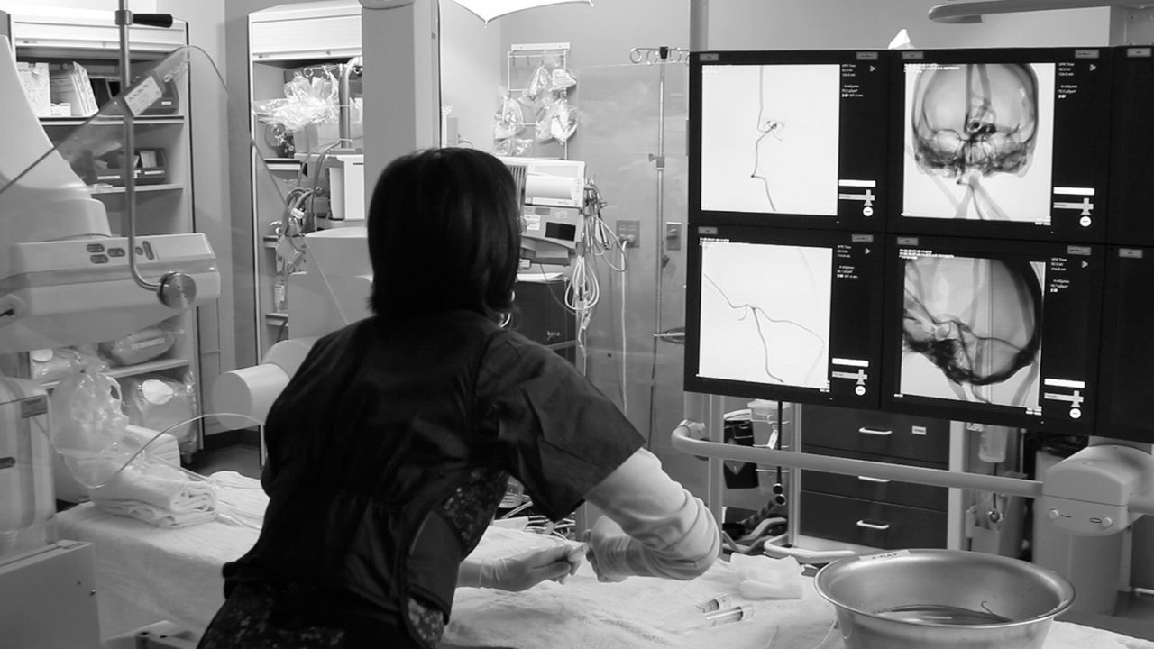
The resident using the simulator can practice all aspects of the procedure at hand. She can practice removing the wire and catheter from the bowl, acquiring the proper fluoro views, using road maps appropriately, and catheter and wire work with actual tactile feedback.

One version of the silicone model is displayed here. This model includes the aortic arch, with carotid and vertebral artery access to the cranial circulation. The anterior cerebral artery, middle cerebral artery, and posterior cerebral artery are represented with customizable aneurysms. Different arch configurations are available or a custom model can be made to match a specific patient's vasculature.

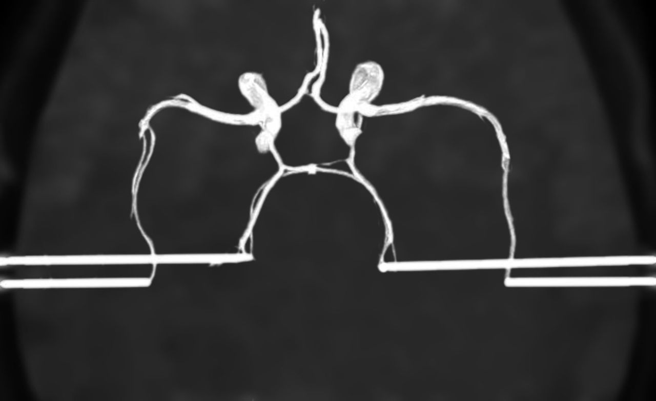
Top down view of a rendition of the intracranial vasculature available in the model. Access vessels have been added, which connect different portions of the cerebral vasculature and allow for recirculation of fluid within the model.
Trainees were tested on the simulator at two different time points. During the first time, trainees received coaching. On the second attempt, they received less coaching, with the trainer interjecting only when the procedure had stalled or the trainee did not know the next step in the procedure. In this case, the trainee's score was decreased.
For each of the two testing sessions, total procedural time and fluoro time were recorded from the biplane fluoro machine. The four subjective categories of navigation, vessel selection, projection setup, and road map usage were scored on a scale from 1 to 5, with 5 being the best score. Scores were assessed by the faculty endovascular neurosurgeon, taking procedural maneuvers, wire technique, and catheter technique into account.
Results
The mean total time to complete a four vessel angiogram improved significantly between sessions 1 and 2 for the 10 trainees. This decreased from 50.7 min in the first session to 42.2 min in the second session (p=0.01). The mean total fluoroscopy time for a four vessel angiogram also decreased significantly, from 11.8 min to 8.8 min (p=0.002).
The four measured technical parameters also improved between the first and second sessions. Average arch navigation scores improved from 3.9 to 4.8 (p=0.029). Vessel selection scores improved significantly from 3.8 to 4.8 (p=0.015). Scores given for setting up projections for the angiographic runs and setting up/using road maps improved from 3.0 to 3.6 and 3.4 to 4.1, respectively. These last two variables, angiographic projection set up (p=0.024) and road map setup/usage (p=0.0095), were also significantly improved (table 1).
Average time and average scores at the first and second testing sessions in the angiography suite with the silicone vascular model
Discussion
Due to the reduced work hours, trainees have to find alternative ways to learn outside of clinical procedures and be more efficient with their inhospital time. Silicone models offer training programs a cost effective means of training residents and fellows to become familiar with the environment of the actual angiography suite. While training on simulators may count towards work hours, it can make each actual case have a higher impact for the trainee and be safer for the patient. Also, they give the residents an opportunity to handle the angiography catheters, wires, and clinical equipment, which they will use when performing a case.
Other simulators, such as the computerized simulators, have several advantages: they are portable, never require radiation for use, and can be loaded with clinical vignettes for trainees to work with. Although computerized simulators have the advantage of being customizable on a case by case basis, they lack the tactile feedback that a silicone model provides when practicing skills such as inserting a shaped catheter or wire into a sheath or navigating the aortic arch. For instance, using a silicone model, a trainee can make a needed adjustment when the wire catches against a vessel wall, responding to the haptic feedback of resistance to advancing the wire. This is not a feature of current computerized simulators.
The other major advantage of the silicone model is that the trainee is required to become facile with the controls of the biplane unit, a skill which is often learned last by trainees. A trainee who is efficient with the controls of the biplane unit will use cones and collimators more effectively and fluoro more sparingly, leading to a lower radiation dose for both the patient and angiographer in the long term. In this study, two practice sessions resulted in shorter fluoro times and better acquisition of road maps and angiographic projections.
Recently it has been proposed that expertise in a certain domain does not depend directly on observed professional experience but on deliberate practice. This theory states that improvement and mastery of a skill requires that a person be cognitively engaged in an activity, which is designed to take place at a specific level of skill and with opportunity for the trainee to pause and rectify errors or be shown a way to improve a certain technique.2 Feedback will often be required for the trainee to understand what aspects of his performance to focus on. While computerized training systems offer the advantage of software to automatically score trainees on objective parameters, a prior study has shown that the only difference between a group of novice subjects and experienced subjects was the fluoroscopy time; the computer could not otherwise tell the difference between the groups.3 This may be because a computer has a difficult time assessing things such as smoothness of movement, decision making, and judgment; there is no replacement for an expert teacher when seeking feedback in these areas. Also, just as a computer generated score seems no substitute for the comments of an experienced mentor, simulators are no replacement for residency and fellowship training and actual patient cases in the biplane fluoro suite.
If exposure to radiation is not desired, then the silicone model can be easily reverted to a videoscopic model, which has been used previously in endovascular training for vascular surgeons and has been shown to be beneficial.4
Additionally, as a potential future direction of this research and training, if a certain case is noteworthy or challenging enough to merit simulator practice devoted to the patient's particular conformation of vasculature, a silicone model can be made to match the patient's vascular anatomy.
As simulators are used more frequently, future work will also include the development of even lower dose fluoroscopy and road mapping protocols to further reduce radiation exposure to the operators.
Overall, our experience with silicone neurosimulators has been extremely positive, with an improvement in trainees’ skills and a reduction in fluoroscopy and procedural time. Experiences with these simulators can improve clinical performance in real patients that allow trainees the opportunity to focus on the more nuanced aspects of a diagnostic or interventional case by getting them comfortable with the more fundamental elements.
Footnotes
-
Contributors BM collected the majority of the data, analyzed the data, and drafted a portion of the manuscript, in addition to approving the final manuscript. CMN collected a portion of the data, analyzed the data, and drafted the majority of the manuscript, critically edited it, and approved the final manuscript. EA collected a portion of the data and assisted in analysis of that data, and approved the final manuscript. KR designed the low dose fluoro protocol used in the study and drafted a portion of the manuscript, and approved the final manuscript. DBN oversaw the project, scored the individual participants, drafted a portion of the manuscript, critically edited it, and approved the final manuscript for submission.
-
Competing interests None.
-
Provenance and peer review Not commissioned; externally peer reviewed.