
Long-COVID and Post-COVID Health Complications: An Up-to-Date Review on Clinical Conditions and Their Possible Molecular Mechanisms

 ,
,  , ,
, ,  , , and
Abstract
:1. Introduction
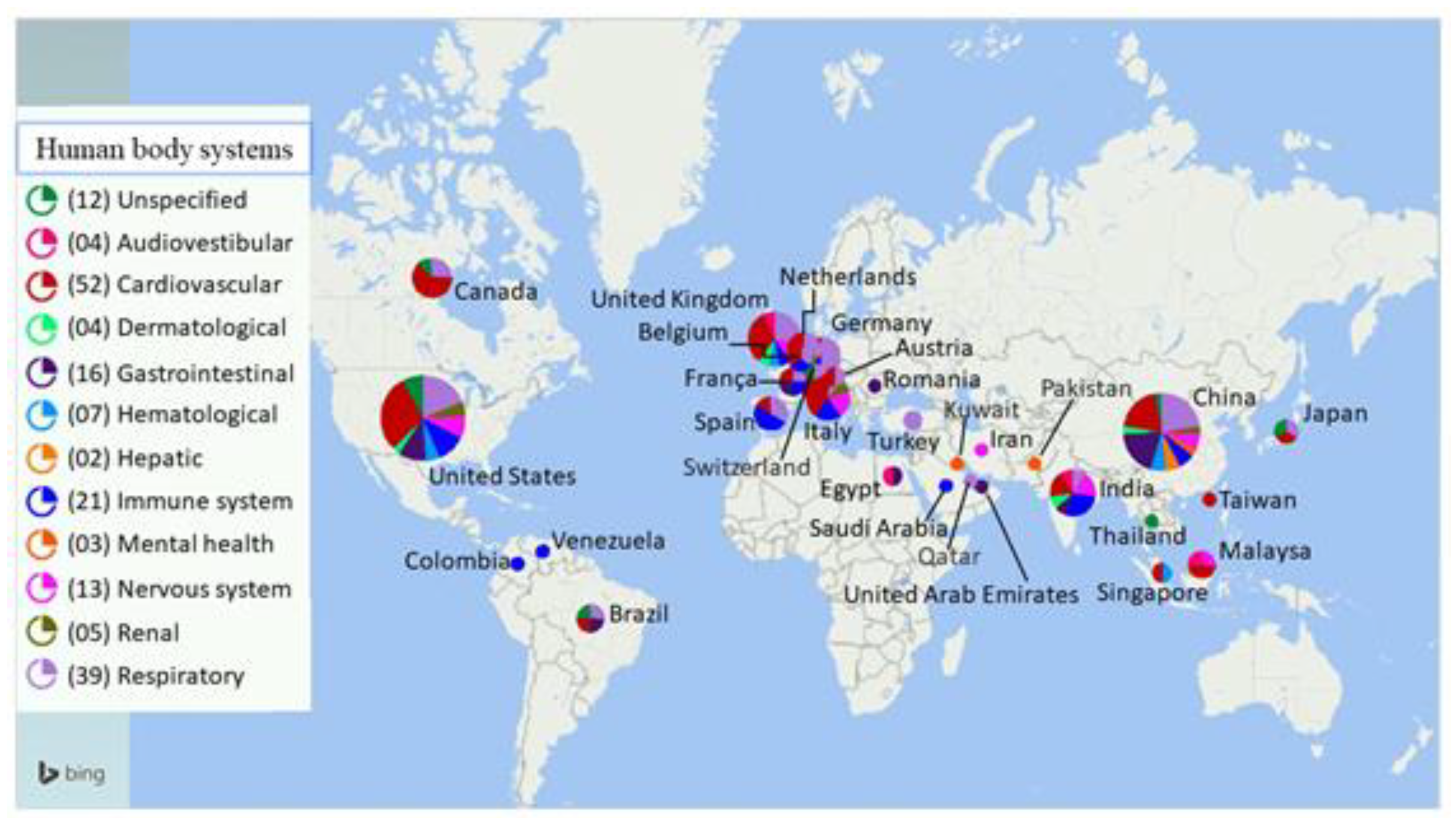
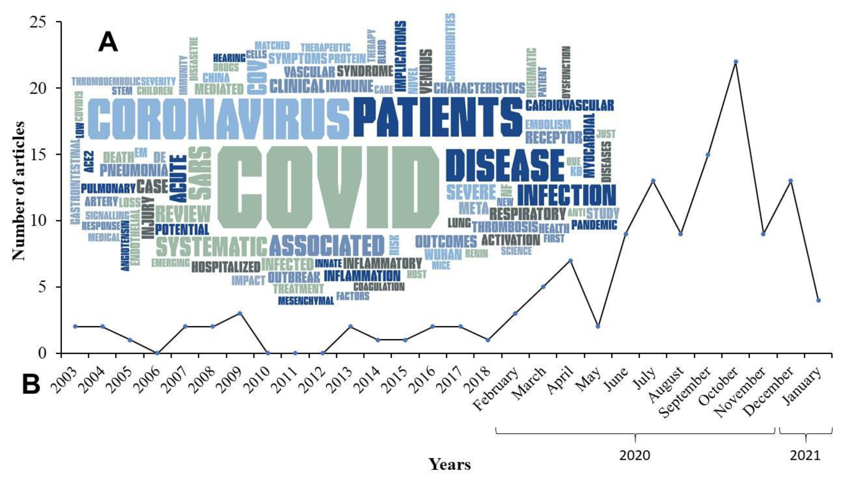
2. Methods
Literature Search Selection Criteria
3. Results
3.1. Complications of the Immune System
Molecular Mechanisms of Immune System Complications
3.2. Complications of the Hematological System
Molecular Mechanisms of Hematological System Complications
3.3. Complications of the Pulmonary System
Molecular Mechanisms of Pulmonary System Complications
3.4. Complications of the Cardiovascular System
Molecular Mechanisms of Cardiovascular System Complications
3.5. Complications of Gastrointestinal, Hepatic, and Renal System
Molecular Mechanisms of Gastrointestinal, Hepatic, and Renal System Complications
3.6. Complications of Skeletomuscular System
Molecular Mechanisms of Skeletomuscular System Complications
3.7. Complications of Nervous System
Molecular Mechanisms of Nervous System Complications
3.8. Complications and Impacts on Mental Health
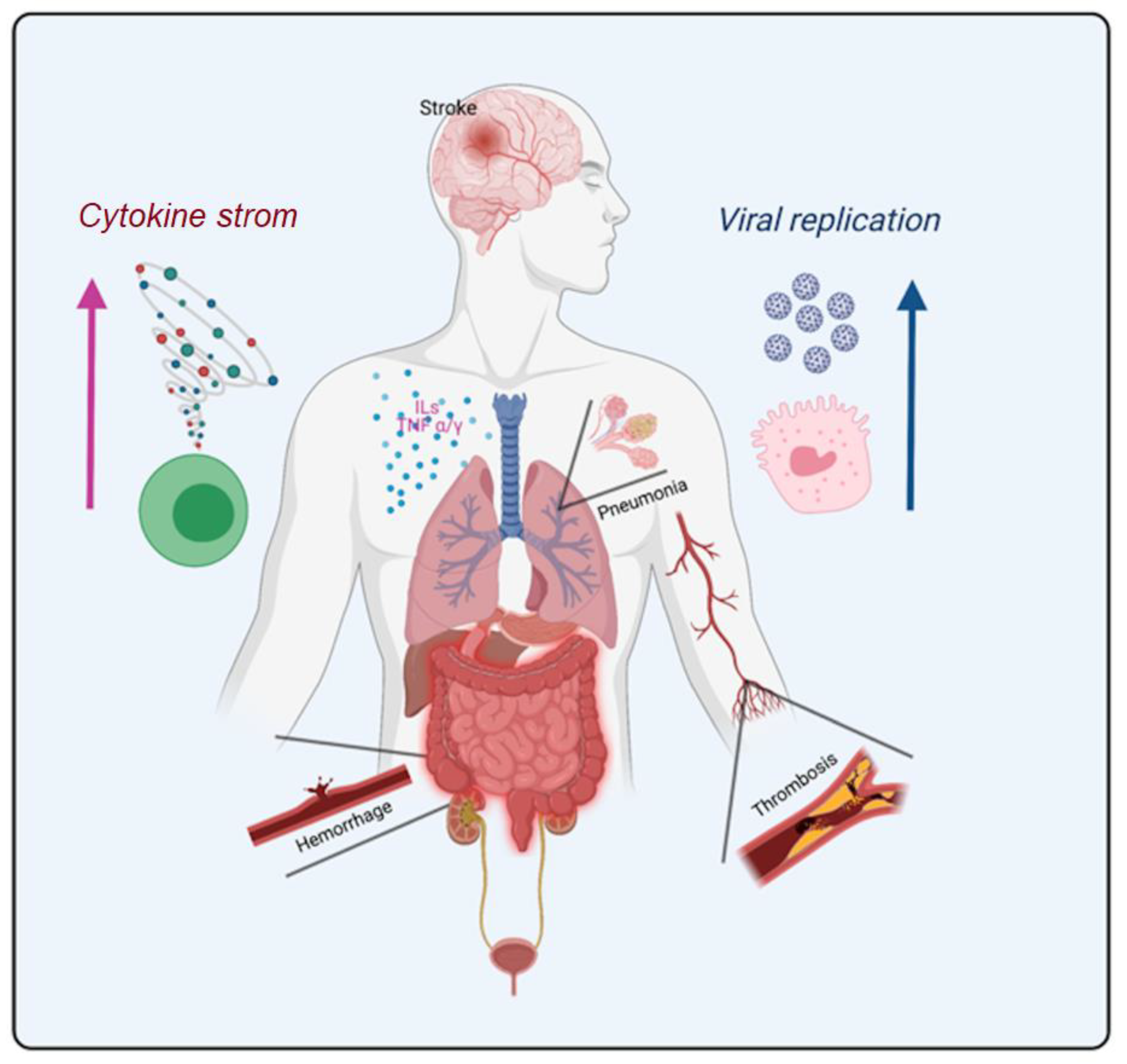
3.9. Is the COVID-19 an Inflammatory Disease Related to Thrombosis?
4. Conclusions and Perspectives
Author Contributions
Funding
Institutional Review Board Statement
Informed Consent Statement
Data Availability Statement
Conflicts of Interest
References
- Zhou, P.; Yang, X.L.; Wang, X.G.; Hu, B.; Zhang, L.; Zhang, W.; Si, H.R.; Zhu, Y.; Li, B.; Huang, C.L.; et al. Addendum: A pneumonia outbreak associated with a new coronavirus of probable bat origin. Nature 2020, 588, E6. [Google Scholar] [CrossRef] [PubMed]
- Fauci, A.S.; Lane, H.C.; Redfield, R.R. Covid-19—Navigating the uncharted. N. Engl. J. Med. 2020, 382, 1268–1269. [Google Scholar] [CrossRef]
- WHO. Coronavirus Disease (Covid-2019) Situation Report—163. 2020. Available online: https://apps.who.int/iris/bitstream/handle/10665/332971/nCoVsitrep01Jul2020-eng.pdf?sequence=1&isAllowed=y (accessed on 1 April 2021).
- Zhu, N.; Zhang, D.; Wang, W.; Li, X.; Yang, B.; Song, J.; Zhao, X.; Huang, B.; Shi, W.; Lu, R.; et al. A novel coronavirus from patients with pneumonia in china, 2019. N. Engl. J. Med. 2020, 382, 727–733. [Google Scholar] [CrossRef]
- Khan, M.; Khan, H.; Khan, S.; Nawaz, M. Epidemiological and clinical characteristics of coronavirus disease (Covid-19) cases at a screening clinic during the early outbreak period: A single-centre study. J. Med. Microbiol. 2020, 69, 1114–1123. [Google Scholar] [CrossRef] [PubMed]
- Pandey, P.; Agarwal, S.; Rajkumar. Lung pathology in Covid-19: A systematic review. Int. J. Appl. Basic Med. Res. 2020, 10, 226–233. [Google Scholar] [CrossRef]
- Guo, T.; Fan, Y.; Chen, M.; Wu, X.; Zhang, L.; He, T.; Wang, H.; Wan, J.; Wang, X.; Lu, Z. Cardiovascular implications of fatal outcomes of patients with coronavirus disease 2019 (Covid-19). JAMA Cardiol. 2020, 5, 811–818. [Google Scholar] [CrossRef] [PubMed] [Green Version]
- Meiler, S.; Hamer, O.W.; Schaible, J.; Zeman, F.; Zorger, N.; Kleine, H.; Rennert, J.; Stroszczynski, C.; Poschenrieder, F. Computed tomography characterization and outcome evaluation of Covid-19 pneumonia complicated by venous thromboembolism. PLoS ONE 2020, 15, e0242475. [Google Scholar] [CrossRef]
- Leung, T.Y.M.; Chan, A.Y.L.; Chan, E.W.; Chan, V.K.Y.; Chui, C.S.L.; Cowling, B.J.; Gao, L.; Ge, M.Q.; Hung, I.F.N.; Ip, M.S.M.; et al. Short- and potential long-term adverse health outcomes of Covid-19: A rapid review. Emerg. Microbes Infect. 2020, 9, 2190–2199. [Google Scholar] [CrossRef]
- Vitiello, A.; Ferrara, F. Pharmacological agents to therapeutic treatment of cardiac injury caused by Covid-19. Life Sci. 2020, 262, 118510. [Google Scholar] [CrossRef] [PubMed]
- Andrade, B.S.; Rangel, F.S.; Santos, N.O.; Freitas, A.D.S.; Soares, W.R.A.; Siqueira, S.; Barh, D.; Goes-Neto, A.; Birbrair, A.; Azevedo, V.A.C. Repurposing approved drugs for guiding Covid-19 prophylaxis: A systematic review. Front. Pharmacol. 2020, 11, 590598. [Google Scholar] [CrossRef]
- Jin, Y.; Ji, W.; Yang, H.; Chen, S.; Zhang, W.; Duan, G. Endothelial activation and dysfunction in Covid-19: From basic mechanisms to potential therapeutic approaches. Signal. Transduct. Target. Ther. 2020, 5, 293. [Google Scholar] [CrossRef]
- Giustino, G.; Pinney, S.P.; Lala, A.; Reddy, V.Y.; Johnston-Cox, H.A.; Mechanick, J.I.; Halperin, J.L.; Fuster, V. Coronavirus and cardiovascular disease, myocardial injury, and arrhythmia: Jacc focus seminar. J. Am. Coll. Cardiol. 2020, 76, 2011–2023. [Google Scholar] [CrossRef] [PubMed]
- Teuwen, L.A.; Geldhof, V.; Pasut, A.; Carmeliet, P. Covid-19: The vasculature unleashed. Nat. Rev. Immunol. 2020, 20, 389–391. [Google Scholar] [CrossRef]
- Grosse, C.; Grosse, A.; Salzer, H.J.F.; Dunser, M.W.; Motz, R.; Langer, R. Analysis of cardiopulmonary findings in Covid-19 fatalities: High incidence of pulmonary artery thrombi and acute suppurative bronchopneumonia. Cardiovasc. Pathol. 2020, 49, 107263. [Google Scholar] [CrossRef] [PubMed]
- Richardson, S.; Hirsch, J.S.; Narasimhan, M.; Crawford, J.M.; McGinn, T.; Davidson, K.W.; the Northwell, C.-R.C.; Barnaby, D.P.; Becker, L.B.; Chelico, J.D.; et al. Presenting characteristics, comorbidities, and outcomes among 5700 patients hospitalized with Covid-19 in the new york city area. JAMA 2020, 323, 2052–2059. [Google Scholar] [CrossRef]
- Najafi, S.; Rajaei, E.; Moallemian, R.; Nokhostin, F. The potential similarities of Covid-19 and autoimmune disease pathogenesis and therapeutic options: New insights approach. Clin. Rheumatol. 2020, 39, 3223–3235. [Google Scholar] [CrossRef]
- Liu, P.P.; Blet, A.; Smyth, D.; Li, H. The science underlying Covid-19: Implications for the cardiovascular system. Circulation 2020, 142, 68–78. [Google Scholar] [CrossRef] [Green Version]
- Palaskas, N.L.; Koutroumpakis, E.; Deswal, A. Covid-19 and cardiovascular health among patients with cancer. Curr. Cardiol. Rep. 2020, 22, 171. [Google Scholar] [CrossRef]
- Escher, R.; Breakey, N.; Lammle, B. Severe Covid-19 infection associated with endothelial activation. Thromb. Res. 2020, 190, 62. [Google Scholar] [CrossRef] [PubMed]
- Varga, Z.; Flammer, A.J.; Steiger, P.; Haberecker, M.; Andermatt, R.; Zinkernagel, A.S.; Mehra, M.R.; Schuepbach, R.A.; Ruschitzka, F.; Moch, H. Endothelial cell infection and endotheliitis in Covid-19. Lancet 2020, 395, 1417–1418. [Google Scholar] [CrossRef]
- Gheblawi, M.; Wang, K.; Viveiros, A.; Nguyen, Q.; Zhong, J.C.; Turner, A.J.; Raizada, M.K.; Grant, M.B.; Oudit, G.Y. Angiotensin-converting enzyme 2: Sars-cov-2 receptor and regulator of the renin-angiotensin system: Celebrating the 20th anniversary of the discovery of ace2. Circ. Res. 2020, 126, 1456–1474. [Google Scholar] [CrossRef] [PubMed]
- Oudit, G.Y.; Kassiri, Z.; Jiang, C.; Liu, P.P.; Poutanen, S.M.; Penninger, J.M.; Butany, J. Sars-coronavirus modulation of myocardial ace2 expression and inflammation in patients with sars. Eur. J. Clin. Investig. 2009, 39, 618–625. [Google Scholar] [CrossRef] [PubMed]
- Hamming, I.; Cooper, M.E.; Haagmans, B.L.; Hooper, N.M.; Korstanje, R.; Osterhaus, A.D.; Timens, W.; Turner, A.J.; Navis, G.; van Goor, H. The emerging role of ace2 in physiology and disease. J. Pathol. 2007, 212, 1–11. [Google Scholar] [CrossRef] [PubMed]
- Chansrichavala, P.; Chantharaksri, U.; Sritara, P.; Chaiyaroj, S.C. Atorvastatin attenuates tlr4-mediated nf-kappab activation in a myd88-dependent pathway. Asian Pac. J. Allergy Immunol. 2009, 27, 49–57. [Google Scholar] [PubMed]
- Sheahan, T.; Morrison, T.E.; Funkhouser, W.; Uematsu, S.; Akira, S.; Baric, R.S.; Heise, M.T. Myd88 is required for protection from lethal infection with a mouse-adapted sars-cov. PLoS Pathog. 2008, 4, e1000240. [Google Scholar] [CrossRef] [Green Version]
- DeDiego, M.L.; Nieto-Torres, J.L.; Regla-Nava, J.A.; Jimenez-Guardeno, J.M.; Fernandez-Delgado, R.; Fett, C.; Castano-Rodriguez, C.; Perlman, S.; Enjuanes, L. Inhibition of nf-kappab-mediated inflammation in severe acute respiratory syndrome coronavirus-infected mice increases survival. J. Virol. 2014, 88, 913–924. [Google Scholar] [CrossRef] [Green Version]
- Rios, J.L. Effects of triterpenes on the immune system. J. Ethnopharmacol. 2010, 128, 1–14. [Google Scholar] [CrossRef]
- Gaestel, M.; Kotlyarov, A.; Kracht, M. Targeting innate immunity protein kinase signalling in inflammation. Nat. Rev. Drug Discov. 2009, 8, 480–499. [Google Scholar] [CrossRef]
- Barh, D.; Tiwari, S.; Andrade, B.S.; Weener, M.E.; Goes-Neto, A.; Azevedo, V.; Ghosh, P.; Blum, K.; Ganguly, N.K. A novel multi-omics-based highly accurate prediction of symptoms, comorbid conditions, and possible long-term complications of Covid-19. Mol. Omics 2021. [Google Scholar] [CrossRef] [PubMed]
- Baas, J.; Schotten, M.; Plume, A.; Côté, G.; Karimi, R. Scopus as a curated, high-quality bibliometric data source for academic research in quantitative science studies. Quant. Sci. Stud. 2020, 1, 377–386. [Google Scholar] [CrossRef]
- Ballew, B.S. Elsevier’s scopus® database. J. Electron. Resour. Med. Librar. 2009, 6, 245–252. [Google Scholar] [CrossRef]
- Canas, C.A. The triggering of post-Covid-19 autoimmunity phenomena could be associated with both transient immunosuppression and an inappropriate form of immune reconstitution in susceptible individuals. Med. Hypotheses 2020, 145, 110345. [Google Scholar] [CrossRef] [PubMed]
- Loarce-Martos, J.; Garcia-Fernandez, A.; Lopez-Gutierrez, F.; Garcia-Garcia, V.; Calvo-Sanz, L.; Del Bosque-Granero, I.; Teran-Tinedo, M.A.; Boteanu, A.; Bachiller-Corral, J.; Vazquez-Diaz, M. High rates of severe disease and death due to sars-cov-2 infection in rheumatic disease patients treated with rituximab: A descriptive study. Rheumatol. Int. 2020, 40, 2015–2021. [Google Scholar] [CrossRef] [PubMed]
- Carrillo-Larco, R.M.; Altez-Fernandez, C.; Ravaglia, S.; Vizcarra, J.A. Covid-19 and guillain-barre syndrome: A systematic review of case reports. Wellcome Open Res. 2020, 5, 107. [Google Scholar] [CrossRef]
- Dhanalakshmi, K.; Venkataraman, A.; Balasubramanian, S.; Madhusudan, M.; Amperayani, S.; Putilibai, S.; Sadasivam, K.; Ramachandran, B.; Ramanan, A.V. Epidemiological and clinical profile of pediatric inflammatory multisystem syndrome—Temporally associated with sars-cov-2 (pims-ts) in indian children. Indian Pediatr. 2020, 57, 1010–1014. [Google Scholar] [CrossRef]
- Sedaghat, Z.; Karimi, N. Guillain barre syndrome associated with Covid-19 infection: A case report. J. Clin. Neurosci. 2020, 76, 233–235. [Google Scholar] [CrossRef]
- Garcia-Manzanedo, S.; Lopez de la Oliva Calvo, L.; Ruiz Alvarez, L. Guillain-barre syndrome after Covid-19 infection. Med. Clin. 2020, 155, 366. [Google Scholar] [CrossRef]
- Ferri, C.; Giuggioli, D.; Raimondo, V.; L’Andolina, M.; Tavoni, A.; Cecchetti, R.; Guiducci, S.; Ursini, F.; Caminiti, M.; Varcasia, G.; et al. Covid-19 and rheumatic autoimmune systemic diseases: Report of a large italian patients series. Clin. Rheumatol. 2020, 39, 3195–3204. [Google Scholar] [CrossRef] [PubMed]
- Veenstra, J.; Buechler, C.R.; Robinson, G.; Chapman, S.; Adelman, M.; Tisack, A.; Dimitrion, P.; Todter, E.; Kohen, L.; Lim, H.W. Antecedent immunosuppressive therapy for immune-mediated inflammatory diseases in the setting of a Covid-19 outbreak. J. Am. Acad. Dermatol. 2020, 83, 1696–1703. [Google Scholar] [CrossRef] [PubMed]
- Pablos, J.L.; Galindo, M.; Carmona, L.; Lledo, A.; Retuerto, M.; Blanco, R.; Gonzalez-Gay, M.A.; Martinez-Lopez, D.; Castrejon, I.; Alvaro-Gracia, J.M.; et al. Clinical outcomes of hospitalised patients with Covid-19 and chronic inflammatory and autoimmune rheumatic diseases: A multicentric matched cohort study. Ann. Rheum. Dis. 2020, 79, 1544–1549. [Google Scholar] [CrossRef]
- Haberman, R.H.; Castillo, R.; Chen, A.; Yan, D.; Ramirez, D.; Sekar, V.; Lesser, R.; Solomon, G.; Neimann, A.L.; Blank, R.B.; et al. Covid-19 in patients with inflammatory arthritis: A prospective study on the effects of comorbidities and disease-modifying antirheumatic drugs on clinical outcomes. Arthritis Rheumatol. 2020, 72, 1981–1989. [Google Scholar] [CrossRef] [PubMed]
- Hassen, L.M.; Almaghlouth, I.A.; Hassen, I.M.; Daghestani, M.H.; Almohisen, A.A.; Alqurtas, E.M.; Alkhalaf, A.; Bedaiwi, M.K.; Omair, M.A.; Almogairen, S.M.; et al. Impact of Covid-19 outbreak on rheumatic patients’ perceptions and behaviors: A cross-sectional study. Int. J. Rheum. Dis. 2020, 23, 1541–1549. [Google Scholar] [CrossRef]
- Sadanandam, A.; Bopp, T.; Dixit, S.; Knapp, D.; Emperumal, C.P.; Vergidis, P.; Rajalingam, K.; Melcher, A.; Kannan, N. A blood transcriptome-based analysis of disease progression, immune regulation, and symptoms in coronavirus-infected patients. Cell Death Discov. 2020, 6, 141. [Google Scholar] [CrossRef]
- Goncalves, L.F.; Gonzales, A.I.; Patatt, F.S.A.; Paiva, K.M.; Haas, P. Kawasaki and Covid-19 disease in children: A systematic review. Rev. Assoc. Med. Bras. 2020, 66 (Suppl. S2), 136–142. [Google Scholar] [CrossRef]
- Shah, S.; Danda, D.; Kavadichanda, C.; Das, S.; Adarsh, M.B.; Negi, V.S. Autoimmune and rheumatic musculoskeletal diseases as a consequence of sars-cov-2 infection and its treatment. Rheumatol. Int. 2020, 40, 1539–1554. [Google Scholar] [CrossRef]
- Zachariah, U.; Nair, S.C.; Goel, A.; Balasubramanian, K.A.; Mackie, I.; Elias, E.; Eapen, C.E. Targeting raised von willebrand factor levels and macrophage activation in severe Covid-19: Consider low volume plasma exchange and low dose steroid. Thromb. Res. 2020, 192, 2. [Google Scholar] [CrossRef]
- Aird, W.C. Phenotypic heterogeneity of the endothelium: Ii. Representative vascular beds. Circ. Res. 2007, 100, 174–190. [Google Scholar] [CrossRef] [Green Version]
- Boisrame-Helms, J.; Kremer, H.; Schini-Kerth, V.; Meziani, F. Endothelial dysfunction in sepsis. Curr. Vasc. Pharmacol. 2013, 11, 150–160. [Google Scholar] [PubMed]
- Cobos-Siles, M.; Cubero-Morais, P.; Arroyo-Jimenez, I.; Rey-Hernandez, M.; Hernandez-Gomez, L.; Vargas-Parra, D.J.; Gonzalez-Fernandez, M.; Cazorla-Gonzalez, M.; Gabella-Martin, M.; Ruiz-Albi, T.; et al. Cause-specific death in hospitalized individuals infected with sars-cov-2: More than just acute respiratory failure or thromboembolic events. Intern. Emerg. Med. 2020, 15, 1533–1544. [Google Scholar] [CrossRef] [PubMed]
- D’Errico, S.; Zanon, M.; Montanaro, M.; Radaelli, D.; Sessa, F.; Di Mizio, G.; Montana, A.; Corrao, S.; Salerno, M.; Pomara, C. More than pneumonia: Distinctive features of sars-cov-2 infection. From autopsy findings to clinical implications: A systematic review. Microorganisms 2020, 8, 1642. [Google Scholar] [CrossRef] [PubMed]
- Shi, S.; Qin, M.; Cai, Y.; Liu, T.; Shen, B.; Yang, F.; Cao, S.; Liu, X.; Xiang, Y.; Zhao, Q.; et al. Characteristics and clinical significance of myocardial injury in patients with severe coronavirus disease 2019. Eur. Heart J. 2020, 41, 2070–2079. [Google Scholar] [CrossRef] [PubMed]
- Peiris, J.S.; Lai, S.T.; Poon, L.L.; Guan, Y.; Yam, L.Y.; Lim, W.; Nicholls, J.; Yee, W.K.; Yan, W.W.; Cheung, M.T.; et al. Coronavirus as a possible cause of severe acute respiratory syndrome. Lancet 2003, 361, 1319–1325. [Google Scholar] [CrossRef] [Green Version]
- Wang, D.; Hu, B.; Hu, C.; Zhu, F.; Liu, X.; Zhang, J.; Wang, B.; Xiang, H.; Cheng, Z.; Xiong, Y.; et al. Clinical characteristics of 138 hospitalized patients with 2019 novel coronavirus-infected pneumonia in wuhan, china. JAMA 2020, 323, 1061–1069. [Google Scholar] [CrossRef] [PubMed]
- Al-Khawaga, S.; Abdelalim, E.M. Potential application of mesenchymal stem cells and their exosomes in lung injury: An emerging therapeutic option for Covid-19 patients. Stem Cell Res. Ther. 2020, 11, 437. [Google Scholar] [CrossRef] [PubMed]
- Piazza, G.; Campia, U.; Hurwitz, S.; Snyder, J.E.; Rizzo, S.M.; Pfeferman, M.B.; Morrison, R.B.; Leiva, O.; Fanikos, J.; Nauffal, V.; et al. Registry of arterial and venous thromboembolic complications in patients with Covid-19. J. Am. Coll. Cardiol. 2020, 76, 2060–2072. [Google Scholar] [CrossRef]
- Sakr, Y.; Giovini, M.; Leone, M.; Pizzilli, G.; Kortgen, A.; Bauer, M.; Tonetti, T.; Duclos, G.; Zieleskiewicz, L.; Buschbeck, S.; et al. Pulmonary embolism in patients with coronavirus disease-2019 (Covid-19) pneumonia: A narrative review. Ann. Intensive Care 2020, 10, 124. [Google Scholar] [CrossRef]
- Sidarta-Oliveira, D.; Jara, C.P.; Ferruzzi, A.J.; Skaf, M.S.; Velander, W.H.; Araujo, E.P.; Velloso, L.A. Sars-cov-2 receptor is co-expressed with elements of the kinin-kallikrein, renin-angiotensin and coagulation systems in alveolar cells. Sci. Rep. 2020, 10, 19522. [Google Scholar] [CrossRef]
- Fujii, H.; Tsuji, T.; Yuba, T.; Tanaka, S.; Suga, Y.; Matsuyama, A.; Omura, A.; Shiotsu, S.; Takumi, C.; Ono, S.; et al. High levels of anti-ssa/ro antibodies in Covid-19 patients with severe respiratory failure: A case-based review: High levels of anti-ssa/ro antibodies in Covid-19. Clin. Rheumatol. 2020, 39, 3171–3175. [Google Scholar] [CrossRef]
- Ooi, M.W.X.; Rajai, A.; Patel, R.; Gerova, N.; Godhamgaonkar, V.; Liong, S.Y. Pulmonary thromboembolic disease in Covid-19 patients on ct pulmonary angiography—Prevalence, pattern of disease and relationship to d-dimer. Eur. J. Radiol. 2020, 132, 109336. [Google Scholar] [CrossRef]
- Uppuluri, E.M.; Shapiro, N.L. Development of pulmonary embolism in a nonhospitalized patient with Covid-19 who did not receive venous thromboembolism prophylaxis. Am. J. Health Syst. Pharm. 2020, 77, 1957–1960. [Google Scholar] [CrossRef]
- Pisano, T.J.; Joki, J.; Hon, B.; Cuccurullo, S. Pulmonary embolism after acute spinal cord injury and Covid-19. Am. J. Phys. Med. Rehabil. 2020, 99, 982–985. [Google Scholar] [CrossRef]
- George, P.M.; Barratt, S.L.; Condliffe, R.; Desai, S.R.; Devaraj, A.; Forrest, I.; Gibbons, M.A.; Hart, N.; Jenkins, R.G.; McAuley, D.F.; et al. Respiratory follow-up of patients with Covid-19 pneumonia. Thorax 2020, 75, 1009–1016. [Google Scholar] [CrossRef] [PubMed]
- Van Kruijsdijk, R.C.; de Jong, P.A.; Abrahams, A.C. Pulmonary vein thrombosis in Covid-19. BMJ Case Rep. 2020, 13, e239986. [Google Scholar] [CrossRef]
- Korkmaz, B.; Lesner, A.; Marchand-Adam, S.; Moss, C.; Jenne, D.E. Lung protection by cathepsin c inhibition: A new hope for Covid-19 and ards? J. Med. Chem. 2020, 63, 13258–13265. [Google Scholar] [CrossRef] [PubMed]
- Pillai, P.; Joseph, J.P.; Fadzillah, N.H.M.; Mahmod, M. Covid-19 and major organ thromboembolism: Manifestations in neurovascular and cardiovascular systems. J. Stroke Cerebrovasc. Dis. 2021, 30, 105427. [Google Scholar] [CrossRef] [PubMed]
- Khan, M.Z.; Jamal, Y.; Sutton, B.; Rauf, F. Venous thromboembolism in patients with Covid-19 and correlation with d-dimers: A single-centre experience. BMJ Open Respir. Res. 2020, 7, e000779. [Google Scholar] [CrossRef] [PubMed]
- Ackermann, M.; Verleden, S.E.; Kuehnel, M.; Haverich, A.; Welte, T.; Laenger, F.; Vanstapel, A.; Werlein, C.; Stark, H.; Tzankov, A.; et al. Pulmonary vascular endothelialitis, thrombosis, and angiogenesis in Covid-19. N. Engl. J. Med. 2020, 383, 120–128. [Google Scholar] [CrossRef] [PubMed]
- Tanaka, T.; Narazaki, M.; Kishimoto, T. Immunotherapeutic implications of il-6 blockade for cytokine storm. Immunotherapy 2016, 8, 959–970. [Google Scholar] [CrossRef]
- Booth, C.M.; Matukas, L.M.; Tomlinson, G.A.; Rachlis, A.R.; Rose, D.B.; Dwosh, H.A.; Walmsley, S.L.; Mazzulli, T.; Avendano, M.; Derkach, P.; et al. Clinical features and short-term outcomes of 144 patients with Sars in the greater Toronto area. JAMA 2003, 289, 2801–2809. [Google Scholar] [CrossRef] [Green Version]
- Badawi, A.; Ryoo, S.G. Prevalence of comorbidities in the middle east respiratory syndrome coronavirus (mers-cov): A systematic review and meta-analysis. Int. J. Infect. Dis. 2016, 49, 129–133. [Google Scholar] [CrossRef] [Green Version]
- Smeeth, L.; Thomas, S.L.; Hall, A.J.; Hubbard, R.; Farrington, P.; Vallance, P. Risk of myocardial infarction and stroke after acute infection or vaccination. N. Engl. J. Med. 2004, 351, 2611–2618. [Google Scholar] [CrossRef] [Green Version]
- Li, B.; Yang, J.; Zhao, F.; Zhi, L.; Wang, X.; Liu, L.; Bi, Z.; Zhao, Y. Prevalence and impact of cardiovascular metabolic diseases on Covid-19 in china. Clin. Res. Cardiol. 2020, 109, 531–538. [Google Scholar] [CrossRef] [PubMed]
- Thygesen, K.; Alpert, J.S.; Jaffe, A.S.; Chaitman, B.R.; Bax, J.J.; Morrow, D.A.; White, H.D.; Executive Group on behalf of the Joint European Society of Cardiology/American College of Cardiology/American Heart Association/World Heart Federation Task Force for the Universal Definition of Myocardial, I. Fourth universal definition of myocardial infarction (2018). Circulation 2018, 138, e618–e651. [Google Scholar] [CrossRef] [PubMed]
- Van de Veerdonk, F.L.; Netea, M.G.; Dinarello, C.A.; Joosten, L.A. Inflammasome activation and il-1beta and il-18 processing during infection. Trends Immunol. 2011, 32, 110–116. [Google Scholar] [CrossRef] [PubMed]
- Vallance, P.; Collier, J.; Bhagat, K. Infection, inflammation, and infarction: Does acute endothelial dysfunction provide a link? Lancet 1997, 349, 1391–1392. [Google Scholar] [CrossRef]
- Bermejo-Martin, J.F.; Almansa, R.; Torres, A.; Gonzalez-Rivera, M.; Kelvin, D.J. Covid-19 as a cardiovascular disease: The potential role of chronic endothelial dysfunction. Cardiovasc. Res. 2020, 116, e132–e133. [Google Scholar] [CrossRef] [PubMed]
- Sarkisian, L.; Saaby, L.; Poulsen, T.S.; Gerke, O.; Jangaard, N.; Hosbond, S.; Diederichsen, A.C.; Thygesen, K.; Mickley, H. Clinical characteristics and outcomes of patients with myocardial infarction, myocardial injury, and nonelevated troponins. Am. J. Med. 2016, 129, 446.e5–446.e21. [Google Scholar] [CrossRef] [PubMed]
- Teixeira, R.; Santos, M.; Gil, V. Covid-19 and cardiovascular comorbidities: An update. Rev. Port. Cardiol. 2020, 39, 417–419. [Google Scholar] [CrossRef]
- Moderato, L.; Monello, A.; Lazzeroni, D.; Binno, S.; Giacalone, R.; Ferraro, S.; Piepoli, M.F.; Villani, G.Q. Takotsubo syndrome during sars-cov-2 pneumonia: A possible cardiovascular complication. G. Ital. Cardiol. 2020, 21, 417–420. [Google Scholar]
- Qin, C.; Zhou, L.; Hu, Z.; Zhang, S.; Yang, S.; Tao, Y.; Xie, C.; Ma, K.; Shang, K.; Wang, W.; et al. Dysregulation of immune response in patients with coronavirus 2019 (Covid-19) in Wuhan, China. Clin. Infect. Dis. 2020, 71, 762–768. [Google Scholar] [CrossRef]
- Hobai, I.A.; Edgecomb, J.; LaBarge, K.; Colucci, W.S. Dysregulation of intracellular calcium transporters in animal models of sepsis-induced cardiomyopathy. Shock 2015, 43, 3–15. [Google Scholar] [CrossRef] [Green Version]
- Reynolds, H.R.; Adhikari, S.; Pulgarin, C.; Troxel, A.B.; Iturrate, E.; Johnson, S.B.; Hausvater, A.; Newman, J.D.; Berger, J.S.; Bangalore, S.; et al. Renin-angiotensin-aldosterone system inhibitors and risk of Covid-19. N. Engl. J. Med. 2020, 382, 2441–2448. [Google Scholar] [CrossRef] [PubMed]
- Scott, A.J.; O’Dea, K.P.; O’Callaghan, D.; Williams, L.; Dokpesi, J.O.; Tatton, L.; Handy, J.M.; Hogg, P.J.; Takata, M. Reactive oxygen species and p38 mitogen-activated protein kinase mediate tumor necrosis factor alpha-converting enzyme (tace/adam-17) activation in primary human monocytes. J. Biol. Chem. 2011, 286, 35466–35476. [Google Scholar] [CrossRef] [PubMed] [Green Version]
- Patel, V.B.; Clarke, N.; Wang, Z.; Fan, D.; Parajuli, N.; Basu, R.; Putko, B.; Kassiri, Z.; Turner, A.J.; Oudit, G.Y. Angiotensin ii induced proteolytic cleavage of myocardial ace2 is mediated by tace/adam-17: A positive feedback mechanism in the ras. J. Mol. Cell Cardiol. 2014, 66, 167–176. [Google Scholar] [CrossRef]
- Tavazzi, G.; Pellegrini, C.; Maurelli, M.; Belliato, M.; Sciutti, F.; Bottazzi, A.; Sepe, P.A.; Resasco, T.; Camporotondo, R.; Bruno, R.; et al. Myocardial localization of coronavirus in Covid-19 cardiogenic shock. Eur. J. Heart Fail. 2020, 22, 911–915. [Google Scholar] [CrossRef] [PubMed] [Green Version]
- Wenzel, P.; Kopp, S.; Gobel, S.; Jansen, T.; Geyer, M.; Hahn, F.; Kreitner, K.F.; Escher, F.; Schultheiss, H.P.; Munzel, T. Evidence of sars-cov-2 mrna in endomyocardial biopsies of patients with clinically suspected myocarditis tested negative for Covid-19 in nasopharyngeal swab. Cardiovasc. Res. 2020, 116, 1661–1663. [Google Scholar] [CrossRef]
- Chen, R.; Yu, Y.L.; Li, W.; Liu, Y.; Lu, J.X.; Chen, F.; Zhou, Q.; Xia, Z.Y.; Gao, L.; Meng, Q.T.; et al. Gastrointestinal symptoms associated with unfavorable prognosis of Covid-19 patients: A retrospective study. Front. Med. 2020, 7, 608259. [Google Scholar] [CrossRef]
- Zhang, L.B.; Pang, R.R.; Qiao, Q.H.; Wang, Z.H.; Xia, X.Y.; Wang, C.J.; Xu, X.L. Successful recovery of Covid-19-associated recurrent diarrhea and gastrointestinal hemorrhage using convalescent plasma. Mil. Med. Res. 2020, 7, 45. [Google Scholar] [CrossRef]
- Zhong, P.; Xu, J.; Yang, D.; Shen, Y.; Wang, L.; Feng, Y.; Du, C.; Song, Y.; Wu, C.; Hu, X.; et al. Covid-19-associated gastrointestinal and liver injury: Clinical features and potential mechanisms. Signal. Transduct. Target. Ther. 2020, 5, 256. [Google Scholar] [CrossRef]
- Bilal, M.; Sawhney, M.S.; Feuerstein, J.D. Coronavirus disease-2019: Implications for the gastroenterologist. Curr. Opin. Gastroenterol. 2021, 37, 23–29. [Google Scholar] [CrossRef]
- Martin, T.A.; Wan, D.W.; Hajifathalian, K.; Tewani, S.; Shah, S.L.; Mehta, A.; Kaplan, A.; Ghosh, G.; Choi, A.J.; Krisko, T.I.; et al. Gastrointestinal bleeding in patients with coronavirus disease 2019: A matched case-control study. Am. J. Gastroenterol. 2020, 115, 1609–1616. [Google Scholar] [CrossRef] [PubMed]
- Abdelmohsen, M.A.; Alkandari, B.M.; Gupta, V.K.; ElBeheiry, A.A. Diagnostic value of abdominal sonography in confirmed Covid-19 intensive care patients. Egypt. J. Radiol. Nucl. Med. 2020, 51, 1–7. [Google Scholar] [CrossRef]
- Villapol, S. Gastrointestinal symptoms associated with Covid-19: Impact on the gut microbiome. Transl. Res. 2020, 226, 57–69. [Google Scholar] [CrossRef]
- Vodnar, D.C.; Mitrea, L.; Teleky, B.E.; Szabo, K.; Calinoiu, L.F.; Nemes, S.A.; Martau, G.A. Coronavirus disease (Covid-19) caused by (sars-cov-2) infections: A real challenge for human gut microbiota. Front. Cell Infect. Microbiol. 2020, 10, 575559. [Google Scholar] [CrossRef]
- Weber, T.K.; Leandro-Merhi, V.A.; Bernasconi, I.; Oliveira, M.R.M. Nutritional therapy in hospital care of in-patients with Covid-19: Evidence, consensus and practice guidelines. Rev. Nutr. 2020, e200212. [Google Scholar] [CrossRef]
- Dong, Z.Y.; Xiang, B.J.; Jiang, M.; Sun, M.J.; Dai, C. The prevalence of gastrointestinal symptoms, abnormal liver function, digestive system disease and liver disease in Covid-19 infection: A systematic review and meta-analysis. J. Clin. Gastroenterol. 2021, 55, 67–76. [Google Scholar] [CrossRef] [PubMed]
- Wang, J.G.; Cui, H.R.; Tang, H.B.; Deng, X.L. Gastrointestinal symptoms and fecal nucleic acid testing of children with 2019 coronavirus disease: A systematic review and meta-analysis. Sci. Rep. 2020, 10, 17846. [Google Scholar] [CrossRef]
- Ye, Q.; Wang, B.; Zhang, T.; Xu, J.; Shang, S. The mechanism and treatment of gastrointestinal symptoms in patients with Covid-19. Am. J. Physiol Gastrointest Liver Physiol 2020, 319, G245–G252. [Google Scholar] [CrossRef]
- Ramanathan, M.; Chueng, T.; Fernandez, E.; Gonzales-Zamora, J. Concomitant renal and splenic infarction as a complication of Covid-19: A case report and literature review. Infez Med. 2020, 28, 611–615. [Google Scholar]
- Acharya, S.; Anwar, S.; Siddiqui, F.S.; Shabih, S.; Manchandani, U.; Dalezman, S. Renal artery thrombosis in Covid-19. IDCases 2020, 22, e00968. [Google Scholar] [CrossRef] [PubMed]
- Idilman, I.S.; Telli Dizman, G.; Ardali Duzgun, S.; Irmak, I.; Karcaaltincaba, M.; Inkaya, A.C.; Demirkazik, F.; Durhan, G.; Gulsun Akpinar, M.; Ariyurek, O.M.; et al. Lung and kidney perfusion deficits diagnosed by dual-energy computed tomography in patients with Covid-19-related systemic microangiopathy. Eur. Radiol. 2021, 31, 1090–1099. [Google Scholar] [CrossRef]
- Trottein, F.; Sokol, H. Potential causes and consequences of gastrointestinal disorders during a sars-cov-2 infection. Cell Rep. 2020, 32, 107915. [Google Scholar] [CrossRef]
- Singh, A.K.; Bhushan, B.; Maurya, A.; Mishra, G.; Singh, S.K.; Awasthi, R. Novel coronavirus disease 2019 (Covid-19) and neurodegenerative disorders. Dermatol. Ther. 2020, 33, e13591. [Google Scholar] [CrossRef]
- Sanghvi, A.R. Covid-19: An overview for dermatologists. Int. J. Dermatol. 2020, 59, 1437–1449. [Google Scholar] [CrossRef] [PubMed]
- Zahedi Niaki, O.; Anadkat, M.J.; Chen, S.T.; Fox, L.P.; Harp, J.; Micheletti, R.G.; Nambudiri, V.E.; Pasieka, H.B.; Shinohara, M.M.; Rosenbach, M.; et al. Navigating immunosuppression in a pandemic: A guide for the dermatologist from the covid task force of the medical dermatology society and society of dermatology hospitalists. J. Am. Acad. Dermatol. 2020, 83, 1150–1159. [Google Scholar] [CrossRef] [PubMed]
- Bonometti, R.; Sacchi, M.C.; Stobbione, P.; Lauritano, E.C.; Tamiazzo, S.; Marchegiani, A.; Novara, E.; Molinaro, E.; Benedetti, I.; Massone, L.; et al. The first case of systemic lupus erythematosus (sle) triggered by Covid-19 infection. Eur. Rev. Med. Pharmacol. Sci. 2020, 24, 9695–9697. [Google Scholar] [PubMed]
- Gokhale, Y.; Patankar, A.; Holla, U.; Shilke, M.; Kalekar, L.; Karnik, N.D.; Bidichandani, K.; Baveja, S.; Joshi, A. Dermatomyositis during Covid-19 pandemic (a case series): Is there a cause effect relationship? J. Assoc. Physicians India 2020, 68, 20–24. [Google Scholar]
- Nigro, E.; Polito, R.; Alfieri, A.; Mancini, A.; Imperlini, E.; Elce, A.; Krustrup, P.; Orru, S.; Buono, P.; Daniele, A. Molecular mechanisms involved in the positive effects of physical activity on coping with Covid-19. Eur. J. Appl. Physiol. 2020, 120, 2569–2582. [Google Scholar] [CrossRef]
- Li, Y.C.; Bai, W.Z.; Hashikawa, T. The neuroinvasive potential of sars-cov2 may play a role in the respiratory failure of Covid-19 patients. J. Med. Virol. 2020, 92, 552–555. [Google Scholar] [CrossRef]
- Disser, N.P.; De Micheli, A.J.; Schonk, M.M.; Konnaris, M.A.; Piacentini, A.N.; Edon, D.L.; Toresdahl, B.G.; Rodeo, S.A.; Casey, E.K.; Mendias, C.L. Musculoskeletal consequences of Covid-19. J. Bone Joint Surg. Am. 2020, 102, 1197–1204. [Google Scholar] [CrossRef]
- Greve, J.M.D.; Brech, G.C.; Quintana, M.; Soares, A.L.S.; Alonso, A.C. Impacts of Covid-19 on the immune, neuromuscular, and musculoskeletal systems and rehabilitation. Rev. Bras. Med. Esporte 2020, 26, 4. [Google Scholar] [CrossRef]
- Townsend, L.; Dyer, A.H.; Jones, K.; Dunne, J.; Mooney, A.; Gaffney, F.; O’Connor, L.; Leavy, D.; O’Brien, K.; Dowds, J.; et al. Persistent fatigue following sars-cov-2 infection is common and independent of severity of initial infection. PLoS ONE 2020, 15, e0240784. [Google Scholar] [CrossRef]
- Lau, H.M.; Lee, E.W.; Wong, C.N.; Ng, G.Y.; Jones, A.Y.; Hui, D.S. The impact of severe acute respiratory syndrome on the physical profile and quality of life. Arch. Phys. Med. Rehabil. 2005, 86, 1134–1140. [Google Scholar] [CrossRef] [Green Version]
- McCray, P.B., Jr.; Pewe, L.; Wohlford-Lenane, C.; Hickey, M.; Manzel, L.; Shi, L.; Netland, J.; Jia, H.P.; Halabi, C.; Sigmund, C.D.; et al. Lethal infection of k18-hace2 mice infected with severe acute respiratory syndrome coronavirus. J. Virol. 2007, 81, 813–821. [Google Scholar] [CrossRef] [PubMed] [Green Version]
- Gumucio, J.P.; Qasawa, A.H.; Ferrara, P.J.; Malik, A.N.; Funai, K.; McDonagh, B.; Mendias, C.L. Reduced mitochondrial lipid oxidation leads to fat accumulation in myosteatosis. FASEB J. 2019, 33, 7863–7881. [Google Scholar] [CrossRef] [PubMed]
- Mendias, C.L.; Roche, S.M.; Harning, J.A.; Davis, M.E.; Lynch, E.B.; Sibilsky Enselman, E.R.; Jacobson, J.A.; Claflin, D.R.; Calve, S.; Bedi, A. Reduced muscle fiber force production and disrupted myofibril architecture in patients with chronic rotator cuff tears. J. Shoulder Elbow Surg. 2015, 24, 111–119. [Google Scholar] [CrossRef] [PubMed]
- Leung, T.W.; Wong, K.S.; Hui, A.C.; To, K.F.; Lai, S.T.; Ng, W.F.; Ng, H.K. Myopathic changes associated with severe acute respiratory syndrome: A postmortem case series. Arch. Neurol. 2005, 62, 1113–1117. [Google Scholar] [CrossRef]
- Mao, L.; Jin, H.; Wang, M.; Hu, Y.; Chen, S.; He, Q.; Chang, J.; Hong, C.; Zhou, Y.; Wang, D.; et al. Neurologic manifestations of hospitalized patients with coronavirus disease 2019 in wuhan, china. JAMA Neurol. 2020, 77, 683–690. [Google Scholar] [CrossRef] [Green Version]
- Favas, T.T.; Dev, P.; Chaurasia, R.N.; Chakravarty, K.; Mishra, R.; Joshi, D.; Mishra, V.N.; Kumar, A.; Singh, V.K.; Pandey, M.; et al. Neurological manifestations of Covid-19: A systematic review and meta-analysis of proportions. Neurol. Sci. 2020, 41, 3437–3470. [Google Scholar] [CrossRef]
- Rhea, E.M.; Logsdon, A.F.; Hansen, K.M.; Williams, L.M.; Reed, M.J.; Baumann, K.K.; Holden, S.J.; Raber, J.; Banks, W.A.; Erickson, M.A. The s1 protein of sars-cov-2 crosses the blood-brain barrier in mice. Nat. Neurosci. 2021, 24, 368–378. [Google Scholar] [CrossRef]
- Saniasiaya, J. Hearing loss in sars-cov-2: What do we know? Ear Nose Throat J. 2021, 100, 152S–154S. [Google Scholar] [CrossRef]
- Samaranayake, L.P.; Fakhruddin, K.S.; Panduwawala, C. Sudden onset, acute loss of taste and smell in coronavirus disease 2019 (Covid-19): A systematic review. Acta Odontol. Scand. 2020, 78, 467–473. [Google Scholar] [CrossRef]
- Almufarrij, I.; Uus, K.; Munro, K.J. Does coronavirus affect the audio-vestibular system? A rapid systematic review. Int. J. Audiol. 2020, 59, 487–491. [Google Scholar] [CrossRef]
- Karimi-Galougahi, M.; Naeini, A.S.; Raad, N.; Mikaniki, N.; Ghorbani, J. Vertigo and hearing loss during the Covid-19 pandemic—Is there an association? Acta Otorhinolaryngol. Ital. 2020, 40, 463–465. [Google Scholar] [CrossRef]
- Salari, M.; Etemadifar, M.; Gharagozli, K.; Etemad, K.; Ashrafi, F.; Ashourizadeh, H. Incidence of anxiety in epilepsy during coronavirus disease (Covid-19) pandemic. Epilepsy Behav. 2020, 112, 107442. [Google Scholar] [CrossRef]
- Monti, G.; Giovannini, G.; Marudi, A.; Bedin, R.; Melegari, A.; Simone, A.M.; Santangelo, M.; Pignatti, A.; Bertellini, E.; Trenti, T.; et al. Anti-nmda receptor encephalitis presenting as new onset refractory status epilepticus in Covid-19. Seizure 2020, 81, 18–20. [Google Scholar] [CrossRef] [PubMed]
- Sripadma, P.V.; Rai, A.; Wadhwa, C. Postpartum atypical posterior reversible encephalopathy syndrome in a Covid-19 patient—An obstetric emergency. J. Stroke Cerebrovasc. Dis. 2020, 29, 105357. [Google Scholar]
- Lee, K.W.; Yusof Khan, A.H.K.; Ching, S.M.; Chia, P.K.; Loh, W.C.; Abdul Rashid, A.M.; Baharin, J.; Inche Mat, L.N.; Wan Sulaiman, W.A.; Devaraj, N.K.; et al. Stroke and novel coronavirus infection in humans: A systematic review and meta-analysis. Front. Neurol. 2020, 11, 579070. [Google Scholar] [CrossRef] [PubMed]
- Tu, T.M.; Goh, C.; Tan, Y.K.; Leow, A.S.; Pang, Y.Z.; Chien, J.; Shafi, H.; Chan, B.P.; Hui, A.; Koh, J.; et al. Cerebral venous thrombosis in patients with Covid-19 infection: A case series and systematic review. J. Stroke Cerebrovasc. Dis. 2020, 29, 105379. [Google Scholar] [CrossRef]
- Yamakawa, M.; Kuno, T.; Mikami, T.; Takagi, H.; Gronseth, G. Clinical characteristics of stroke with Covid-19: A systematic review and meta-analysis. J. Stroke Cerebrovasc. Dis. 2020, 29, 105288. [Google Scholar] [CrossRef]
- Frisullo, G.; Bellavia, S.; Scala, I.; Piano, C.; Morosetti, R.; Brunetti, V.; Calabresi, P.; Della Marca, G. Stroke and covid19: Not only a large-vessel disease. J. Stroke Cerebrovasc. Dis. 2020, 29, 105074. [Google Scholar] [CrossRef]
- Dakay, K.; Cooper, J.; Bloomfield, J.; Overby, P.; Mayer, S.A.; Nuoman, R.; Sahni, R.; Gulko, E.; Kaur, G.; Santarelli, J.; et al. Cerebral venous sinus thrombosis in Covid-19 infection: A case series and review of the literature. J. Stroke Cerebrovasc. Dis. 2021, 30, 105434. [Google Scholar] [CrossRef]
- Asif, R.; O’Mahony, M.S. Rare complication of Covid-19 presenting as isolated headache. BMJ Case Rep. 2020, 13, e239275. [Google Scholar] [CrossRef]
- Paliogiannis, P.; Mangoni, A.A.; Dettori, P.; Nasrallah, G.K.; Pintus, G.; Zinellu, A. D-dimer concentrations and Covid-19 severity: A systematic review and meta-analysis. Front. Public Health 2020, 8, 432. [Google Scholar] [CrossRef]
- Bareeqa, S.B.; Ahmed, S.I.; Samar, S.S.; Yasin, W.; Zehra, S.; Monese, G.M.; Gouthro, R.V. Prevalence of depression, anxiety and stress in china during Covid-19 pandemic: A systematic review with meta-analysis. Int. J. Psychiatry Med. 2020. [Google Scholar] [CrossRef] [PubMed]
- Burhamah, W.; AlKhayyat, A.; Oroszlanyova, M.; AlKenane, A.; Almansouri, A.; Behbehani, M.; Karimi, N.; Jafar, H.; AlSuwaidan, M. The psychological burden of the Covid-19 pandemic and associated lockdown measures: Experience from 4000 participants. J. Affect. Disord. 2020, 277, 977–985. [Google Scholar] [CrossRef] [PubMed]
- Luo, M.; Guo, L.; Yu, M.; Jiang, W.; Wang, H. The psychological and mental impact of coronavirus disease 2019 (Covid-19) on medical staff and general public—A systematic review and meta-analysis. Psychiatry Res. 2020, 291, 113190. [Google Scholar] [CrossRef] [PubMed]
- Silva, D.F.O.; Cobucci, R.N.; Soares-Rachetti, V.P.; Lima, S.; Andrade, F.B. Prevalence of anxiety among health professionals in times of Covid-19: A systematic review with meta-analysis. Cien Saude Colet 2021, 26, 693–710. [Google Scholar] [CrossRef]
- Tang, N.; Li, D.; Wang, X.; Sun, Z. Abnormal coagulation parameters are associated with poor prognosis in patients with novel coronavirus pneumonia. J. Thromb. Haemost. 2020, 18, 844–847. [Google Scholar] [CrossRef] [Green Version]
- Thachil, J. The versatile heparin in Covid-19. J. Thromb. Haemost. 2020, 18, 1020–1022. [Google Scholar] [CrossRef] [PubMed] [Green Version]
- Vinayagam, S.; Sattu, K. Sars-cov-2 and coagulation disorders in different organs. Life Sci. 2020, 260, 118431. [Google Scholar] [CrossRef]
- Iba, T.; Connors, J.M.; Levy, J.H. The coagulopathy, endotheliopathy, and vasculitis of Covid-19. Inflamm. Res. 2020, 69, 1181–1189. [Google Scholar] [CrossRef] [PubMed]
- Vernuccio, F.; Lombardo, F.P.; Cannella, R.; Panzuto, F.; Giambelluca, D.; Arzanauskaite, M.; Midiri, M.; Cabassa, P. Thromboembolic complications of Covid-19: The combined effect of a pro-coagulant pattern and an endothelial thrombo-inflammatory syndrome. Clin. Radiol. 2020, 75, 804–810. [Google Scholar] [CrossRef] [PubMed]
- Ma, J.; Bai, J. Protective effects of heparin on endothelial cells in sepsis. Int. J. Clin. Exp. Med. 2015, 8, 5547–5552. [Google Scholar] [PubMed]
- Liu, Y.; Mu, S.; Li, X.; Liang, Y.; Wang, L.; Ma, X. Unfractionated heparin alleviates sepsis-induced acute lung injury by protecting tight junctions. J. Surg. Res. 2019, 238, 175–185. [Google Scholar] [CrossRef]
- Young, E. The anti-inflammatory effects of heparin and related compounds. Thromb. Res. 2008, 122, 743–752. [Google Scholar] [CrossRef]
- Li, J.P.; Vlodavsky, I. Heparin, heparan sulfate and heparanase in inflammatory reactions. Thromb. Haemost. 2009, 102, 823–828. [Google Scholar]
- Esmon, C.T. Targeting factor xa and thrombin: Impact on coagulation and beyond. Thromb. Haemost. 2014, 111, 625–633. [Google Scholar] [CrossRef]
- Poterucha, T.J.; Libby, P.; Goldhaber, S.Z. More than an anticoagulant: Do heparins have direct anti-inflammatory effects? Thromb. Haemost. 2017, 117, 437–444. [Google Scholar] [CrossRef]
- Mousavi, S.; Moradi, M.; Khorshidahmad, T.; Motamedi, M. Anti-inflammatory effects of heparin and its derivatives: A systematic review. Adv. Pharmacol. Sci. 2015, 2015, 507151. [Google Scholar] [CrossRef] [Green Version]
- Martínez, M.M.L.; Contreras, M.A.; Marin, W.; D’Marco, L. Statins in Covid-19: Is there any foundation? Clin. Investig. Arterioscler. 2020, 32, 278–281. [Google Scholar]
- Xu, L.; Liu, J.; Lu, M.; Yang, D.; Zheng, X. Liver injury during highly pathogenic human coronavirus infections. Liver Int. 2020, 40, 998–1004. [Google Scholar] [CrossRef] [PubMed] [Green Version]
- Newman, C.B.; Preiss, D.; Tobert, J.A.; Jacobson, T.A.; Page, R.L., 2nd; Goldstein, L.B.; Chin, C.; Tannock, L.R.; Miller, M.; Raghuveer, G.; et al. Statin safety and associated adverse events: A scientific statement from the american heart association. Arterioscler. Thromb. Vasc. Biol. 2019, 39, e38–e81. [Google Scholar] [CrossRef] [Green Version]
- Lala, A.; Johnson, K.W.; Januzzi, J.L.; Russak, A.J.; Paranjpe, I.; Richter, F.; Zhao, S.; Somani, S.; Van Vleck, T.; Vaid, A.; et al. Prevalence and impact of myocardial injury in patients hospitalized with Covid-19 infection. J. Am. Coll. Cardiol. 2020, 76, 533–546. [Google Scholar] [CrossRef] [PubMed]
- Chen, X.; Yan, L.; Fei, Y.; Zhang, C. Laboratory abnormalities and risk factors associated with in-hospital death in patients with severe Covid-19. J. Clin. Lab. Anal. 2020, 34, e23467. [Google Scholar] [CrossRef] [PubMed]
- Bavishi, C.; Bonow, R.O.; Trivedi, V.; Abbott, J.D.; Messerli, F.H.; Bhatt, D.L. Special article—Acute myocardial injury in patients hospitalized with Covid-19 infection: A review. Prog. Cardiovasc. Dis. 2020, 63, 682–689. [Google Scholar] [CrossRef] [PubMed]


| 1 Keywords/Strings | 2 Number of Recovered Articles |
|---|---|
| TITLE-ABS-KEY (Covid-19 OR sars-cov-2 OR 2019-n-cov AND “autoimmune disease” OR “rheumatoid arthritis” OR “toxic shock syndrome” OR thromboembolism) AND (LIMIT-TO (DOCTYPE, “ar”) OR LIMIT-TO (DOCTYPE, “re”)) | 973 |
| TITLE-ABS-KEY (Covid-19 OR sars-cov-2 OR 2019-n-cov AND “intravascular disseminated coagulation” OR ICD OR “pulmonary embolism” OR “Postviral pulmonary fibrosis” OR “pulmonary thromboembolism” OR “cardiac arrhythmia” OR “heart failure” OR “gastrointestinal symptoms” OR “gastrointestinal complications” OR “Orroetic olfactory dysfunction” OR ortorium “Dysfunction taste” OR stroke OR stroke OR GBS OR encephalitis OR encephalopathy OR “ischemic stroke” OR “intracerebral haemorrhage” OR “cerebrovascular disease”) | 125 |
| TITLE-ABS-KEY (Covid-19 OR sars-cov-2 OR 2019-n-cov AND “pulmonary thromboembolism” OR “cardiac arrhythmia” OR “heart failure” OR “gastrointestinal symptoms” OR encephalopathy) AND (LIMIT-TO (DOCTYPE, “ar”) OR LIMIT-TO (DOCTYPE, “re”)) | 1398 |
| TITLE-ABS-KEY (Covid-19 OR sars-cov-2 OR 2019-n-cov AND ‘erythematous AND rash’ OR “macular rash” OR “papular rash” OR “maculopapular rash “ OR “ pseudo-chilblain lesions “ OR “ vesicular lesions “ OR livedo OR necrosis OR “ oral ulcers “ OR blisters OR “ herpetiform lesions “ OR “skin rash” OR “post-traumatic stress disorder “ OR ptsd OR depression OR “ anxiety disorder “) | 20 |
| TITLE-ABS-KEY (Covid-19 OR sars-cov-2 OR 2019-n-cov AND insomnia OR “impaired attention” OR anxiety OR “impaired memory” OR mood OR depression OR “depressive disorders” OR “anxiety disorder” “somatoform pain disorder” OR “panic disorder” OR “Chronic fatigue” OR “autism spectrum disorder” OR “attention deficit disorder” OR HRT OR hyperactivity) | 65 |
| System | Symptoms | Post-COVID | Long-COVID |
|---|---|---|---|
| Audio vestibular | Ageusia | X | |
| Anosmia | X | ||
| Hyposmia | X | ||
| Hearing Loss | X | ||
| Cardiovascular | Myocarditis | X | |
| Heart Failure | X | ||
| Myocardial Hypertrophy | X | ||
| Mild to severe coronary artery atherosclerosis | X | X | |
| Focal myocardial fibrosis | X | ||
| Acute myocardial infarction Type I | X | ||
| Acute myocardial infarction Type II | X | ||
| Cardiogenic shock | X | ||
| Arrhythmia | X | ||
| Pericardial disease | X | ||
| Takotsubo syndrome | X | ||
| Chronic heart disease | X | ||
| Severe coronary artery | X | ||
| Dermatological | Psoriasis | X | |
| Systemic Lupus Erythematosus | X | ||
| Vasculitis | X | ||
| Dermatomyositis | X | ||
| Chronic rheumatological disease | X | ||
| Gastrointestinal | Diarrhea | X | |
| Nausea | X | ||
| Vomit | X | ||
| Abdominal pain | X | ||
| Anorexia | X | ||
| Acid reflux | X | ||
| Gastrointestinal bleeding | X | ||
| Lack of appetite | X | ||
| Constipation | X | ||
| Changes in the lung-intestine-brain axis | X | ||
| Changes in the intestinal flora | X | ||
| Disorders and disintegration of intestinal microorganisms | X | ||
| Microbiota dysbiosis | X | ||
| Dysfunction of intestinal metabolites | X | ||
| Hematological | Breakdown of hemostasis | X | |
| Endoteliitis | X | ||
| Disseminated intravascular coagulation | X | ||
| Prothrombotic phenotype | X | ||
| Coagulative disease | X | ||
| Hepatic | Alteration of inflammatory biomarkers of liver damage | X | |
| Macro and micro thromboembolic | X | ||
| Immune system | Secondary autoimmune symptoms associated with immunosuppression | X | |
| Vascular inflammation and myocarditis | X | ||
| Guillain-Barret syndrome | X | ||
| Motor paralysis | X | ||
| Rheumatoid arthritis | X | ||
| Arthralgia | X | ||
| Myalgia | X | ||
| Weakness | X | ||
| Kawazaki disease | X | ||
| Mental Health | Depression | X | |
| Panic syndrome | X | ||
| Anxiety | X | ||
| Stress | X | ||
| Psychiatric disorders | X | ||
| Anguish | X | ||
| Insomnia | X | ||
| Negative psychosocial effects | X | ||
| Panic Syndrome | X | ||
| Nervous system | Headaches | X | |
| Spasms | X | ||
| Convulsions | X | ||
| Confusion | X | ||
| Visual impairment | X | ||
| Nerve pain | X | ||
| Dizziness | X | ||
| Conscience problems | X | ||
| Nausea | X | ||
| Vomiting | X | ||
| Hemiplegia | X | ||
| Ataxia | X | ||
| Stroke (AVC) | X | ||
| Cerebral hemorrhage | X | ||
| Nonspecific neurological symptoms | |||
| Epileptic seizures | X | ||
| Myalgia | X | ||
| Anti-N-Methyl-D-Aspartate encephalitis (rNMDA) | X | ||
| Atypical postpartum reversible encephalopathy syndrome | X | ||
| Renal | Renal disfunction | X | |
| Renal systemic microangiopathy with micro-thrombosis | X | ||
| Pulmonary | Pulmonary infarction | X | |
| Pulmonary Hemorrhage | X | ||
| Respiratory failure | X | ||
| Pulmonary thromboembolism | X | ||
| Pulmonary embolism | X | ||
| Pneumonia | X | ||
| Secondary bronchopneumonia | X | X | |
| Pulmonary vein thrombosis | X | ||
| Post-viral pulmonary fibrosis | X | X | |
| Chronic respiratory failure | X | X | |
| Dyspnea | X | ||
| Cough | X | ||
| Chest pain | X | ||
| Hemptysis | X | ||
| Skeletomuscular | Dermatomyositis | X | |
| Generalized weakness | X | ||
| Fatigue | X | ||
| Muscle fiber atrophy | X | ||
| Extensive myalgia | X | ||
| Muscle dysfunction | X | ||
| Deficit in muscle strength and endurance | X | ||
| Generalized muscle atrophy | X | ||
| Sporadic and focal necrosis of muscle fibers | X | ||
| Neuronal demyelination | X |
Publisher’s Note: MDPI stays neutral with regard to jurisdictional claims in published maps and institutional affiliations. |
© 2021 by the authors. Licensee MDPI, Basel, Switzerland. This article is an open access article distributed under the terms and conditions of the Creative Commons Attribution (CC BY) license (https://creativecommons.org/licenses/by/4.0/).
Share and Cite
Silva Andrade, B.; Siqueira, S.; de Assis Soares, W.R.; de Souza Rangel, F.; Santos, N.O.; dos Santos Freitas, A.; Ribeiro da Silveira, P.; Tiwari, S.; Alzahrani, K.J.; Góes-Neto, A.; et al. Long-COVID and Post-COVID Health Complications: An Up-to-Date Review on Clinical Conditions and Their Possible Molecular Mechanisms. Viruses 2021, 13, 700. https://doi.org/10.3390/v13040700
Silva Andrade B, Siqueira S, de Assis Soares WR, de Souza Rangel F, Santos NO, dos Santos Freitas A, Ribeiro da Silveira P, Tiwari S, Alzahrani KJ, Góes-Neto A, et al. Long-COVID and Post-COVID Health Complications: An Up-to-Date Review on Clinical Conditions and Their Possible Molecular Mechanisms. Viruses. 2021; 13(4):700. https://doi.org/10.3390/v13040700
Chicago/Turabian StyleSilva Andrade, Bruno, Sérgio Siqueira, Wagner Rodrigues de Assis Soares, Fernanda de Souza Rangel, Naiane Oliveira Santos, Andria dos Santos Freitas, Priscila Ribeiro da Silveira, Sandeep Tiwari, Khalid J Alzahrani, Aristóteles Góes-Neto, and et al. 2021. "Long-COVID and Post-COVID Health Complications: An Up-to-Date Review on Clinical Conditions and Their Possible Molecular Mechanisms" Viruses 13, no. 4: 700. https://doi.org/10.3390/v13040700