
ABSTRACT
Background Prolonged past exposure to secondhand tobacco smoke (SHS) is associated with exercise limitation. Pulmonary factors including air trapping contribute to this limitation but the contribution of cardiovascular factors is unclear.
Methods To determine contribution of cardiovascular mechanisms to SHS-associated exercise limitation, we examined the cardiovascular responses to maximum effort exercise testing in 166 never-smokers with remote but prolonged occupational exposure to SHS and no known history of cardiovascular disease except nine with medically-controlled hypertension. We estimated the contribution of oxygen-pulse (proxy for cardiac stroke volume) and changes in systolic (SBP) and diastolic blood pressures (DBP) and heart rate (HR) over workload towards exercise capacity, and examined whether the association of SHS with exercise capacity was mediated through these variables.
Results Oxygen consumption (VO2Peak) and oxygen-pulse (O2-PulsePeak) at peak exercise were 1,516±431mL/min (100±23 %predicted) and 10.6±2.8mL/beat (117±25 %predicted), respectively, with 91 (55%) and 43 (26%) of subjects not being able to achieve their maximum predicted values. Sixty-two percent showed hypertensive response to exercise by at least one established criterion. In adjusted models, VO2Peak was associated directly with O2-Pulse and inversely with rise of SBP and DBP over workload (all P<0.05). Moreover, SHS exposure association with VO2Peak was mainly (84%) mediated through its effect on oxygen-pulse (P=0.034). Notably, although not statistically significant, a large proportion (60%) of air trapping effect on VO2Peak seemed to be mediated through oxygen-pulse (P=0.066).
Discussion In a never-smoker population with remote prolonged exposure to SHS, abnormal escalation of afterload and an SHS-associated reduction in cardiac output contributed to lower exercise capacity.
Introduction
Secondhand tobacco smoke (SHS) remains a major public health problem.1, 2 Although SHS exposure among nonsmokers in the United States has declined from 88% in 1988 to 25% in 2014, the rate of decline has plateaued with one in four nonsmokers, including 14 million children, continue to be exposed to SHS annually between 2011 and 2014.3 This substantial continued exposure is important particularly as the generations that endured the highest amount of SHS exposure are aging, a process which could accentuate the SHS-related health problems that may have been previously too subtle to be recognized.
Although immediate health effects of exposure to SHS have been studied,4–9 its long-term consequences, particularly the effect of remote exposures, have been more difficult to examine in part due to challenges with exposure assessment. Never-smoking flight crew who worked on commercial aircrafts before the enactment of the smoking ban were exposed to heavy SHS in aircraft cabin for many years, in a range similar to the nicotine exposure burden experienced by “light” smokers.10, 11 The regularity of this intense exposure in the cabin work environment lends itself to relatively accurate SHS exposure quantification through employment history,12 and makes the exposed flight crew a unique population in which the long-term health effects of previous exposure to SHS could be examined as a form of “natural” experiment that is also generalizable to other SHS-exposed populations.
In previous studies of never-smoking flight crew with history of remote but prolonged exposure to SHS in aircraft cabin, we examined the pulmonary health effects of long-term exposure to SHS and showed the association of those outcomes with the number of years during which the flight crew were exposed to SHS in aircraft cabin.13–15 While this never-smoking SHS- exposed cohort had no evidence of spirometric chronic obstructive pulmonary disease (COPD) (a preserved ratio of forced expiratory volume in 1 second to forced vital capacity [FEV1/FVC]), they had abnormal lung function that were suggestive of presence of an occult early/mild obstructive lung disease.15 In the current study, we wished to examine the cardiovascular health effects of remote but prolonged exposure to SHS in this never-smoking cohort with evidence of early obstructive lung disease. We hypothesized that prolonged exposure to SHS contributes to exercise limitation through its adverse cardiovascular health effects, in addition to its contribution through pulmonary mechanisms (air trapping). To evaluate this hypothesis, we analyzed the cohort’s cardiovascular response to maximum effort cardiopulmonary exercise testing.
Methods
Study Overview
This was a post-hoc analysis of data collected as part of the Secondhand Smoke Respiratory Health Study, an observational cohort study of nonsmoking subjects with a range of occupational SHS exposure, as previously described.13–15 Briefly, between July 2007 and July 2015, we recruited US airline flight crewmembers with a history of occupational exposure to SHS, along with nonsmoker controls without such occupational exposure, who were participating in a larger study of cardiopulmonary health effects of prolonged remote exposure to SHS.15 The participants were characterized by respiratory symptom questionnaires, full pulmonary function testing (PFT), and a maximum effort cardiopulmonary exercise testing (CPET). We used the data from this cohort to perform a post-hoc analysis to determine the associations among exercise capacity (volume of oxygen uptake at peak exercise [VO2Peak]), cardiovascular responses to maximum effort CPET (oxygen-pulse [O2-Pulse; a proxy for cardiac stroke volume], systolic and diastolic blood pressure [SBP and DBP], and heart rate [HR]), years of airline employment during which the subjects worked in smoky cabin (cabin SHS exposure), and their interactions with each other as well as with air trapping (RV/TLC), which we had previously shown to be associated with VO2Peak.
Study Population
The Secondhand Smoke Respiratory Health Study recruited the US airline flight crewmembers as part of an investigation of the potential adverse health effects of the cabin environment on those employed before and after introduction of the ban on smoking in U.S. commercial aircraft. Crewmembers were eligible to participate in the study if they had worked ≥5 years in aircraft. A referent group of “sea-level” subjects who lived in San Francisco Bay area and had never been employed as airline crewmembers were also recruited. All subjects were nonsmokers defined by never-smoking or, in ever smokers, no smoking for ≥20 years and a cumulative history of smoking <20 pack-years. Subjects with known history of pulmonary (such as asthma or COPD) or cardiovascular disease (such as coronary artery disease or heart failure) were excluded. Subjects with known history of hypertension were included if their blood pressure was medically controlled as defined by SBP≤150 and DBP≤90 at the time of visit. Overall, out of the 179 subjects who underwent cardiopulmonary exercise testing (CPET), 166 subjects were included in this analysis.15
The UCSF Institutional Review Board (IRB) and the San Francisco VA Medical Center Committee on Research and Development approved study protocols. Written IRB-approved informed consent and Health Insurance Portability and Accountability Act (HIPAA) were obtained from all study participants. Subjects received monetary compensation for their participation in the study.
SHS Exposure Characterization
SHS exposure was characterized by a questionnaire developed by the UCSF Flight Attendant Medical Research Institute (FAMRI) Center of Excellence,16 and modified to acquire information on airline-related occupational history, as described previously.13, 14 Briefly, this included employer airlines, duration of employment, and flight routes with quantification of “cabin SHS exposure” as the number of years during which the crewmembers were exposed to SHS in aircraft. Other possible sources of SHS exposure were also explored by questioning subjects about their non-cabin exposures in additional settings, as described previously.17
Pulmonary Function Testing
Pulmonary function studies were conducted according to the American Thoracic Society (ATS) and European Respiratory Society (ERS) guidelines18–23. Routine pulmonary function tests were performed in the seated position using a model Vmax 229 CareFusion (CareFusion Corp., Yorba Linda, CA) and nSpire body plethysmograph (nSpire Health Inc., Longmont, CO) as described previously.13 This included measurement spirometry including flows low lung volumes24; lung volume by single breath dilution25, 26 and plethysmography27; airway resistance during panting at functional residual capacity (FRC)28, 29; and single breath carbon monoxide diffusing capacity.30 Bronchodilator responsiveness was not performed. Hyperinflation and air trapping, which is inferred from an increase in FRC or residual volume (RV), was quantified using FRC or RV to total lung capacity (TLC) ratio (FRC/TLC or RV/TLC).
Cardiopulmonary Exercise Testing
Subjects performed physician-supervised, symptom-limited, progressively increasing exercise tests in the supine position on an electromagnetically braked, supine cycle ergometer (Medical Positioning Inc. Kansas City, MO). The protocol consisted of 3-min rest, 1-min unloaded (freewheeling) cycling at 60 to 65 revolution per minute (rpm), followed by increasing work rate of 20 to 40 Watts at 2-minute intervals (stages) to a maximum tolerated, and 5-min of recovery. Subjects were encouraged to give their best effort, and during testing, were encouraged to continue exercise until a VO2 plateau effect on a breath-to-breath analysis of oxygen consumption was visually observed; however, they were advised that they could stop voluntarily at any time they believed they could not continue.
Twelve-lead electrocardiogram (ECG), heart rate (HR), and oxyhemoglobin saturation by pulse oximetry (O2sat) were monitored continuously, and blood pressure (BP) (measured manually by a physician with a cuff) was recorded every 2 min during the second minute of each stage. Minute ventilation (VE), oxygen uptake (VO2), and carbon dioxide output (VCO2) were measured breath-by-breath with an open-circuit metabolic cart (model Vmax 229, CareFusion, Yorba Linda, CA). Immediately before all tests, the gas analyzers were calibrated using reference gases of known concentrations and the ventilometer was calibrated using a 3-liter syringe (Hans Rudolph, Kansas, MO). The metabolic system was verified using four trained technicians who provided monthly exercise values as biological standards for the Laboratory.
Peak exercise gas exchange variables (VO2, VCO2, and VE) were estimated as the last 30- second average value obtained during the highest stage of the exercise test that subject was able to complete, as defined by continuous cycling at the required 60 to 65 rpm during that stage for greater than 1 minute. The volumes of the flow meter, mouthpiece, and filter (70 mL x breathing frequency) were subtracted from VE for the VE/VCO2 calculations. Anaerobic threshold (AT) was determined by the V-slope method.31, 32
The subject’s perception of level of effort and exertion, breathlessness, and fatigue was documented during the second minute of every stage using the modified Borg Rating of Perceived Exertion (Borg), the Category-Ratio Scale anchored at number 10 (CR10).33
Respiratory Symptom Scoring
Respiratory symptoms were assessed using modified Medical Research Council (mMRC) Dyspnea Scale34 and another self-reported questionnaire (UCSF FAMRI Center of Excellence questionnaire16) that elicited symptoms of dyspnea, cough, and subjects’ perception of a decreased level of exertion compared to peers over the year preceding enrollment. A dichotomous indicator of respiratory symptoms was defined by mMRC ≥1 or report of at least one respiratory symptom on the FAMRI questionnaire.
Data Analysis
Percent predicted as well as lower and upper limits of normal (LLN and ULN) values for measures of Spirometry and lung volumes at rest and cardiopulmonary responses to exercise were calculated using Global Lung Function Initiative (GLI), Quanjer et al., and Wasserman predicted formulas, repectively.35–37
Distributions of subject characteristics, pulmonary function, cardiopulmonary exercise, and SHS exposure quantification variables were examined. Changes in HR, SBP, DBP, and O2- Pulse with respect to the workload were approximated by estimating the slopes from linear regression modeling of the measures over workload at each stage. Peak cardiopulmonary exercise variables were estimated using the last 30-second average values obtained during the highest stage of the exercise test as described above.
Associations between exercise capacity (VO2Peak) and each of the cardiovascular outputs (including SBP, DBP, and O2-Pulse) were examined in univariate linear regression models with adjustment for covariates (age, sex, height, and BMI unless noted otherwise) and the baseline value of the respected variable. Similarly, associations between presence of respiratory symptoms (mMRC) and each of the cardiovascular outputs were examined in univariate logistic regression models with adjustment for covariates. Additionally, associations between each of the cardiovascular outputs (SBP, DBP, and O2-Pulse) and baseline air trapping (FRC/TLC or RV/TLC) as a pulmonary factor affecting exercise capacity and cardiovascular outputs,38–41 and SHS exposure were examined using linear regression models.
The cardiovascular variables that were statistically significant predictor of exercise capacity (VO2Peak) (or respiratory symptoms [mMRC]) were then used in comprehensive regression models with inclusion of all relevant cardiovascular outputs, air trapping (FRC/TLC or RV/TLC), and SHS exposure as predictors to examine their association with VO2Peak and mMRC while accounting for the interrelationships among the predictors.
To assess whether associations between VO2Peak (or mMRC) and SHS exposure or air trapping (FRC/TLC or RV/TLC) were mediated through cardiovascular outputs, we performed mediation analyses with VO2Peak (or mMRC) (dependent variable), SHS exposure or air trapping (independent variable), and cardiovascular outputs (mediator variables), with inclusion of covariates using the “mediation” package in R.42 To assess robust estimations, the bootstrap method with 1000 resamples from the data was applied to obtain the estimated proportion of mediation and the corresponding P-value.
For each analysis, the total number of subjects who had complete set of data for that analysis was reported along with the results from the regression modeling or mediation analysis.
RESULTS
Subject Characteristics
The characteristics of the 166 participants included in this analysis are shown in Table 1. The subjects were predominantly women (150 [90%]). All were never-smokers (<100 cigarettes lifetime) with the exception of one who had smoked 0.7 pack-years. All subjects had exposure to SHS with 63% and 35% reporting childhood and adult home SHS exposure, respectively. Ninety (54%) had been exposed to cabin SHS during their airline employment for an average of 16.8±8.8 years (median [IQR] <total range> of 18 [10-24] <1-31> years). None of the subjects included in the analysis reported any history of cardiovascular or pulmonary diseases with the exception of nine who had a history of well-controlled hypertension, eight of whom on medical management (two on lisinopril, one on verapamil, one on atenolol, one on hydrochlorothiazide, one on combination hydrochlorothiazide and triamterene, one on combination hydrochlorothiazide and benazepril, and one on an unknown medication).
All subjects had preserved spirometry (normal FEV1/FVC and FEV1 by LLN criterion) but some had mildly reduced diffusing capacity at 79±10 (median [IQR] <total range> of 79 [72- 86]) <53-101> %predicted). The subjects had FRC/TLC of 0.53±0.08 (at 99±14 %predicted; median [IQR] <total range> of 99 [91-107] <54-129> %predicted) and RV/TLC of 0.34±0.07 (at 90±12 % predicted; median [IQR] <total range> of 90 [82-97] <53-120> % predicted).
Pulmonary response to exercise
Although the average volume of oxygen consumption at peak exercise (VO2Peak) was 1,513±429 mL/min (100±23 %predicted), only 75 (45.2%) of subjects achieved their 100% predicted value (median [IQR] <total range> VO2Peak of 97 [84-113] <53-186> %predicted). The pulmonary response to exercise was remarkable for a peak-exercise minute ventilation (VEPeak) of 56.1±15.0 L/min at 114±26 %predicted that was inappropriately elevated for VO2Peak achieved. Despite this high VEPeak, tidal volumes at peak exercise (VTPeak) only reached a high value of 85% of predicted value (1.68±0.41 L; median [IQR] <total range> of 83 [75-94] <49- 134> %predicted), implicating an abnormally elevated respiratory rate, although the peak-exercise respiratory rate (RRPeak) remained <50 breaths per minute threshold in 89% (148 out of 166) of the subjects and <60 breaths per minute in all but 1 subject (35.0±9.0 breaths/min; median [IQR] <total range> of 33.0 [28.8-40.0] <15.0-69.0> breaths/min) (Table 2). Finally, anaerobic threshold (VO2AT) determined by V slope method was reached at 65±16% of maximum predicted VO2 (median [IQR] <total range> of 66 [53-75] <27-106> %predicted) (Table 2).
Cardiovascular response to exercise
None of the subjects reported any chest pain, chest tightness, lightheadedness, or dizziness during the CPET. None had any clinically significant electrocardiographic (ECG) changes or arrythmia besides occasional premature ventricular contractions that did not increase in frequency with exercise testing. All subjects surpassed their anaerobic threshold. All reported ceasing exercise due to dyspnea or leg fatigue. Nevertheless, many had abnormal cardiovascular response to exercise as described below.
Although only 9 (5%) subjects had reported history of hypertension (with their hypertension well-controlled), 62% percent of the subjects (98 out of 158) showed hypertensive response to exercise by at least one established criterion (Table 2). Oxygen-pulse at peak exercise (O2-PulsePeak) was 10.6±2.8 mL/beat (117±25 %predicted; median [IQR] <total range> of 112 [99-134] <74-189> %predicted), with 43 (25.9%) subjects not achieving their 100% predicted values. At anaerobic threshold, O2-Pulse was 8.7±2.6 mL/beat (73±26 %predicted; median [IQR] <total range> of 71 [55-88] <21-185> %predicted) (Table 2).
Furthermore, ventilatory efficiency (VE/VCO2) at peak exercise and anaerobic threshold were 30.9±4.1 (median [IQR] <total range> of 106 [103-117] <89-144> %predicted) and 31.6±3.9 (median [IQR] <total range> of 110 [105-119] <89-157> %predicted), respectively. The lowest VE/VCO2 was 29.4±4.5 (median [IQR] <total range> of 105 [99-110] <4-207> %predicted) (Table 2).
Association of VO2Peak with cardiovascular response measures, air trapping, and SHS exposure
In univariate models that included each cardiopulmonary exercise testing (CPET) output separately as a predictor with adjustment for age, sex, height, BMI, and baseline value of that CPET variable, VO2Peak was directly associated with O2-Pulse and heart rate at peak exercise (O2-PulsePeak and HRPeak) and inversely with the rate of change in HR, SBP, and DBP over workload (HRSlope, SBPSlope, DBPSlope) (all P<0.05) (Table 3). Similarly, VO2Peak was associated with respiratory rate, tidal volume, and minute ventilation at peak exercise (RRPeak, VTPeak, VEPeak) (all P<0.05) (Table 3). VO2Peak was also inversely associated with measures of air trapping (FRC/TLC and RV/TLC) and with years of exposure to cabin SHS (all P<0.05), but was not significantly associated with non-cabin SHS exposure (Table 3).
In age, sex, height, and BMI-adjusted multivariate modeling that included all cardiovascular outputs of exercise that were significantly associated with respiratory symptoms and VO2Peak in univariate modeling, VO2Peak was directly associated with O2-PulsePeak and HRSlope, and inversely with SBPSlope (Table 4). In multivariate model that included all cardiovascular and pulmonary outputs of exercise that were significantly associated with VO2Peak in univariate modeling, only O2-PulsePeak, HRSlope, SBPSlope, and VEPeak retained their statistical significance (Table 5).
Association of respiratory symptoms with cardiovascular response measures
The associations of respiratory symptoms at rest (as measured by mMRC and FAMRI questionnaires) and at peak exercise (as measured by Borg Scale) from univariate regression modeling with each cardiopulmonary exercise testing (CPET) output after adjustment for age, sex, height, BMI, and the baseline value of each CPET variable examined are shown in Table 6. Overall, respiratory symptoms showed significant associations with only a few CPET output including O2-Pulse, HR, and VE at peak exercise and RV/TLC at baseline. Higher O2-PulsePeak was associated with lower respiratory symptoms at baseline and at peak exercise. On the other hand, higher VEPeak was associated with lower respiratory symptoms at baseline but higher respiratory symptoms at peak exercise.
In age, sex, height, and BMI-adjusted multivariate modeling that included cardiovascular outputs of exercise that were significantly associated with respiratory symptoms and VO2Peak from univariate modeling, only the association of O2-PulsePeak with mMRC maintained its significance such that higher O2-PulsePeak was significantly associated with the lower likelihood of having an mMRC ≥1 (average of 35% decrease in odds ratio [OR] for each 1 mL/beat increase in O2-PulsePeak; P=0.042) (Table 4).
In age, sex, height, and BMI-adjusted multivariate modeling that included all cardiovascular and pulmonary outputs of exercise that were significantly associated with respiratory symptoms and VO2Peak from univariate modeling, no variable was significantly associated with respiratory symptoms, although VEPeak did showed a similar trend of association with respiratory symptoms at rest (direct association with ≥1 mMRC) and during exercise (inverse with Borg Scale) as in univariate modeling (P<0.06) (Table 5).
Association of cardiovascular response measures with SHS exposure
In univariate modeling with adjustment for age, sex, height, and BMI, years cabin SHS exposure was significantly associated with lower exercise capacity as measured by VO2Peak and higher respiratory symptoms as measured by FAMRI questionnaire (Tables 3 and 6). In multivariate modeling with inclusion of all cardiovascular outputs, or all cardiovascular and pulmonary outputs, cabin SHS exposure was only significantly associated with higher respiratory symptoms at rest as measured by FAMRI questionnaire (average of 5% and 6% increase in odds ratio [OR] of reporting respiratory symptoms for each one year increase in cabin SHS exposure; P≤0.018) (Tables 4 and 5).
Among the cardiovascular exercise predictors of VO2Peak, only O2-PulsePeak was significantly associated with years of exposure to cabin SHS, showing a parameter estimate of - 0.036±0.018 (P=0.043), which indicates a decrease in O2-PulsePeak of 0.36 mL/beat for every 10 years of exposure to cabin SHS (Table 7). Mediation analysis showed that cabin SHS exposure association with VO2Peak was mainly (84%) mediated through SHS effect on O2-Pulse (P=0.034) (Table 8).
Cardiovascular response measures and baseline air trapping
Given the physical location of lung, heart, and great vasculature within thoracic cavity, we examined the potential interaction of baseline air trapping and cardiovascular outputs of exercise. To do this, we examined whether the cardiovascular predictors of exercise capacity including O2-Pulse (proxy of stroke volume) and SBPSlope mediate the association of air trapping (FRC/TLC and RV/TLC) with VO2Peak. These mediation analyses showed a marginally non- significant association of O2-PulsePeak with FRC/TLC and RV/TLC (all P>0.05 and <0.13), which suggested that approximately 60% and 52% of air trapping (FRC/TLC and RV/TLC) effect on VO2Peak could be mediated through their effect on stroke volume (O2-Pulse) (Table 9).
Discussion
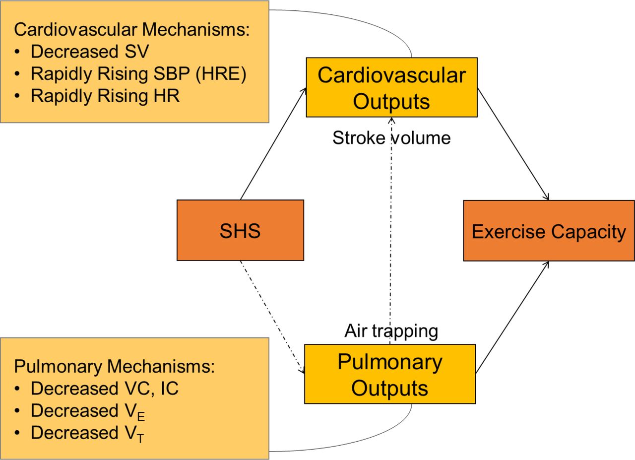
In this observational study of a never-smoking cohort with a history of remote but prolonged exposure to SHS, we found the cohort to have an abnormal cardiovascular response to exercise that was proportional to their SHS exposure. Volume of oxygen uptake at peak exercise (VO2Peak) was associated with years of exposure to SHS, and over 80% of that association was dependent on O2-PulsePeak, suggesting that the effect of SHS exposure on exercise capacity to be mainly mediated through SHS effect on stroke volume and cardiac output. We also found suggestive evidence, although not statistically significant (P=0.066), that pulmonary air trapping (elevated FRC/TLC and RV/TLC) contribute to lower exercise capacity through its effect on O2- PulsePeak, implicating an interacting lung and heart pathophysiology between pulmonary hyperinflation and cardiac output that further impairs exercise capacity (Figure 2). Furthermore, we found over 60% of the participants to have a hypertensive response to exercise, suggesting that abnormal escalation of afterload contributed to lower exercise capacity in this SHS-exposed cohort in whom, with the exception of a few (5.4%) with well-controlled hypertension, none had any known cardiovascular disease.

Abbreviations- SHS=secondhand tobacco smoke; PFT= pulmonary function test; FEV1= forced expiratory volume in 1 second; FVC= forced vital capacity; BMI= body mass index; VO2Max = volume of maximum oxygen uptake.

Illustration of mediation effects between SHS exposure and exercise capacity. Abbreviations- SV= cardiac stroke volume; HRE= hypertensive response to exercise; HR= heart rate; SHS=secondhand tobacco smoke; VC= vital capacity; IC= inspiratory capacity; VE = minute ventilation; VT = volume of tidal breathing.
Overall, we found evidence that prolonged exposure to SHS, even when remote, is associated with cardiovascular abnormalities suggestive of occult cardiovascular dysfunction with potential additional contribution from pulmonary hyperinflation. These abnormalities reveal subtle but lower cardiopulmonary functional reserve, manifested here as lower exercise capacity, and implicate a reduced efficiency of body’s oxygen delivery machinery, which could be disadvantageous during the times of increased cardiopulmonary output demands as in physiological distress or disease.
In previous studies, we have shown this cohort of never-smokers with history of prolonged remote SHS exposure to have abnormal lung function at rest and abnormal pulmonary response to exercise including (1) reduced diffusing capacity at rest,13 (2) reduced pulmonary capillary recruitment (as measured by impaired rise in diffusing capacity) during exercise,14 (3) decreased small airways airflow indices on spirometry (maximal flow in mid- and end-expiratory airflows [FEF25-75% and FEF75%]),13 (4) plethysmographic and radiographic evidence of pulmonary air trapping at rest,15 and (5) progressive (dynamic) pulmonary hyperinflation during exercise.15 Overall, these abnormalities are suggestive of presence of an unrecognized early or mild obstructive lung disease that, while not meeting the spirometric definition of COPD, is consistent with an early/mild disease that could be categorized as “pre-COPD” and could contribute to lower pulmonary reserve and potential adverse health outcomes.43, 44
In the current study, we sought to determine whether there are also subtle cardiovascular abnormalities in this SHS-exposed cohort that are indicative of an occult and/or subclinical cardiovascular disease. Our finding that past exposure to SHS is a predictor of exercise capacity in an O2-pulse-dependent (a proxy of stroke volume and cardiac output) manner is novel and suggests that SHS exposure has lasting effect on cardiac function that is observable years after the exposure has ceased, and is consistent with recent findings that long-standing direct tobacco smoking impairs cardiac systolic function as evident by an increase in left and right ventricle end-systolic volumes and reduced left and right ejection fractions.45 Although it remains unclear how exposure to tobacco smoke, direct or indirect, causes an impairment in cardiac function, an interaction between pulmonary and cardiovascular systems, which occupy the same body cavity (thorax), has been proposed to play a role. Air trapping as measured by lung volumes (FRC/TLC and RV/TLC) is the earliest manifestation of COPD 43, 44 and is associated with reduced exercise capacity due to ventilatory limitation caused by progressive air trapping and pulmonary hyperinflation.15 Changes in lung volumes due to pulmonary hyperinflation could cause increased intra-thoracic pressures, particularly during exertion, and thus adversely affect the cardiovascular function.38, 46 To investigate this possible mechanism, we examined whether air trapping did contribute to exercise capacity through an interaction with cardiac output by performing a mediation analysis among pulmonary air trapping (FRC/TLC and RV/TLC), cardiac stroke volume (O2-pulse), and exercise capacity (VO2Peak) (Figure 2). Although the analysis did not reach statistical significance (P=0.066 and P=0.122 for FRC/TLC and RV/TLC, respectively) and thus could not provide any further corroborating evidence for our hypothesis, the analysis did suggest that a large proportion (60%) of air trapping effect on VO2Peak may be mediated through stroke volume. Further research in this area would be needed to better understand the interaction of cardiovascular and pulmonary systems in the context of exposure to direct or indirect smoke.
Another remarkable observation in our study was the common presence of hypertensive response to exercise (HRE). Despite some data to the contrary,47, 48 studies have described HRE in individuals with or without presence of baseline hypertension to be associated with impaired systolic and/or diastolic dysfunction even in the setting of having negative stress test and preserved left ventricular ejection fraction.49, 50 In fact, increasing evidence indicates that HRE is associated with functional and structural heart abnormalities, future development of hypertension, and increased cardiovascular morbidity and mortality.51–53 In our study, we found a substantial number of participants (62%) to have HRE despite all either having no history of hypertension (95%) or having hypertension that was well-controlled (5%). Studies have reported the prevalence of HRE to be between 3% to 40% among apparently healthy cohorts of varying age, sex, and ethnicity.54–56 Although we did not observe a significant association between presence of HRE and history of SHS exposure, the high prevalence of HRE among this never-smoking but SHS-exposed healthy cohort is remarkable, and suggests a potential pathogenetic role for SHS exposure in development of HRE.
Limitations
Our study has limitations that should be kept in view. First, there may be concerns about the generalizability of the findings because the cohort studied are mostly women, which reflects the demographics of those who worked in airlines as flight crew in the latter half of the last century when smoking in aircraft cabin was permitted. The choice to study the flight crew allowed for overcoming the challenge of long-term SHS exposure assessment by providing the ability to generate a more objective and reproducible exposure index based on employment history and the smoking ban timeline on domestic and international flights of different airlines.13 Women have been reported to be more susceptible to adverse health effects of tobacco smoke.57 However, at the minimum, the findings should be generalizable to women. Second, the cardiovascular findings reported in this study are mainly derived from CPET with no imaging (such as echocardiography and magnetic resonance imaging [MRI]) or other clinical studies to provide additional robust evidence to corroborate our findings. Such studies are needed and are in progress (ClinicalTrials.gov Identifier: NCT04715568). Nevertheless, studies describing cardiovascular health effects of direct smoking using echocardiography and MRI have been previously reported, which corroborate our findings.58, 59 Our report however is the first report that describes the chronic and long-term cardiovascular health effects due to past prolonged exposure to SHS. Third, while we found association of exercise capacity (VO2Peak) with years of SHS exposure, the association of respiratory symptoms with SHS was less striking and less consistent across the different questionnaire platforms. However, it is not uncommon to see differences in scores across different respiratory questionnaires,60 and similarly, baseline respiratory symptoms (mMRC and FAMRI questionnaires) may measure different things and thus produce different scores compared to those done at peak exercise (Borg Scale). For example, subjects who had impairments at baseline and thus were more symptomatic are likely to not perform as well during the exercise and thus may report lesser symptoms in a sub-maximal effort exercise test.
Conclusion
Healthy never-smokers with history of remote but prolonged exposure to SHS have an abnormal cardiovascular response to exercise, which is characterized by a stroke volume (oxygen-pulse) and thus an exercise capacity that are reduced proportional to their years of exposure to SHS. The mechanisms by which past exposure to SHS may limit stroke volume and thus exercise capacity is unknown, but pulmonary hyperinflation may have a contributory effect beyond its role in ventilatory limitation. Furthermore, these SHS-exposed individuals have a considerably higher prevalence of hypertensive response to exercise than that reported in healthy general population. Overall, the abnormal cardiovascular response to exercise in this population reveals the presence of an occult or subclinical pathology that impairs the cardiopulmonary functional reserve and reduces the efficiency of body’s oxygen delivery machinery, which could be disadvantageous during the times of increased cardiopulmonary output demands as in physiological distress or disease.
Data Availability
Data
Acknowledgement
The authors would like to thank Wendy Ching, BS, Charlotte Hunt, BS, and Warren M Gold, MD, for their assistance with performing the cardiopulmonary exercise testing.
Footnotes
Authors’ Contributions Conceived and designed the current manuscript study: MA Developed study protocols: WMG, MA Collected samples and data: SZ, MD, JB, WMG, MA Analyzed and interpreted data: SZ, MD, JB, JK, MA Prepared and edited the manuscript: SZ, MD, JB, WMG, MA Obtained funding: MA
Summary conflict of interest: Authors report no conflict of interest related to this work
Footnote This work was supported by:
Flight Attendant Medical Research Institute (FAMRI) (012500WG and CIA190001 to MA).
California Tobacco-Related Disease Research Program (TRDRP) (T29IR0715 to MA).
The Department of Veterans Affairs (CXV-00125 to MA).
National Library of Medicine Training Grant (NIH: T15LM007442 to SZ)
The funders had no role in study design, data collection and analysis, decision to publish, or preparation of the manuscript. The statements and conclusions in this publication are those of the authors and not necessarily those of the funding agency. The mention of commercial products, their source, or their use in connection with the material reported herein is not to be construed as an actual or implied endorsement of such products.
Notation of prior abstract publication/presentation: Some of the results of this study have been previously reported in the form of an abstract (American Thoracic Society International Meeting 2020: Am J Respir Crit Care Med 2020;201:A7591)
- Abbreviation list:
- COPD
- Chronic obstructive pulmonary disease
- CT
- Computerized tomography
- DM
- Density mask
- EELV
- End-expiratory lung volume
- EFL
- Expiratory flow limitation
- FAMRI
- Flight Attendant Medical Research Institute
- FEF25-75
- Forced expiratory flow at 25% to 75% of FVC
- FEF75
- Forced expiratory flow at 75% of FVC
- FEV1
- Forced expiratory volume in 1 second
- FVC
- Forced vital capacity
- GLI
- Global Lung Function Initiative
- GOLD
- Global Initiative on Obstructive Lung Disease
- HU
- Hounsfield unit
- IC
- Inspiratory capacity
- LLN
- Lower limit of normal
- MEF
- Maximum expiratory flow
- PFT
- Pulmonary function test
- RV
- Residual volume
- SHS
- Secondhand tobacco smoke
- TLC
- Total lung capacity
- VE
- Minute ventilation
- VFL
- Volume of the tidal breath that is flow-limited on expiration
- VO2Max
- Maximum oxygen uptake
- VT
- Tidal volume
- WattsMax
- Maximum work achieved in watts
REFERENCES


